Long-term results of new deproteinized bovine bone material in a maxillary sinus graft procedure
- Affiliations
-
- 1Department Periodontology, Kyung Hee University School of Dentistry, Seoul, Korea.
- 2Department of Dentistry, CHA Bundang Medical Center, CHA University, Seongnam, Korea.
- 3Department Periodontology, Seoul National University School of Dentistry, Seoul, Korea. yjseol@snu.ac.kr
- 4Department of Periodontology, Dankook University Dental Hospital, Cheonan, Korea.
- KMID: 2027824
- DOI: http://doi.org/10.5051/jpis.2014.44.5.259
Abstract
- PURPOSE
The aim of this case report is to present the longitudinal results of sinus grafting using a new demineralized bovine bone material (DBBM) in human cases.
METHODS
A patient with a resorbed maxilla was treated by maxillary sinus grafting using a new deproteinized bovine bone material. After a healing period of 6.5 months, three implants were placed and restored. The patient was periodically recalled and followed up for 5 years after restoration.
RESULTS
Twelve partially edentulous patients (average age, 55.7 years) were followed up. All patients had insufficient residual height in their maxillary posterior area and underwent maxillary sinus graft surgery to increase the height of their maxilla. In all, 27 fixtures were placed in the augmented bone area. On average, 8.6 months later, implants were loaded using provisional or final restorations. The observation period ranged from 27 to 75 months (average, 43.3 months), and the patients did not show any severe resorption of the graft material or any infection during this time.
CONCLUSIONS
Our results show that the new DBBM is useful for a maxillary sinus graft procedure. Good healing responses as well as reliable results were obtained for an average follow-up period of 43.3 months.
MeSH Terms
Figure
-
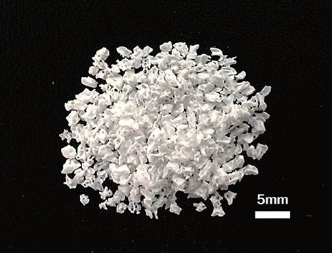
Figure 1 New demineralized bovine bone material particles (particle size, 1-2 mm; original magnification, ×2).
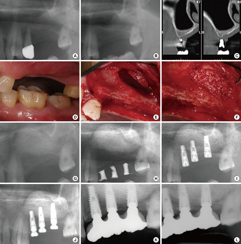
Figure 2 Clinical and radiographic images of patient 5. (A) Panoramic view at the first visit of the patient. (B) Panoramic view before the maxillary sinus graft procedure. The left second premolar was extracted before surgery. (C) Cross-sectional computerized tomography image of the #26 edentulous area. (D) Clinical view on the buccal side of the edentulous area. (E) Window opening of the lateral wall of the left sinus. (F) Demineralized bovine bone material grafting through the window opening. (G) Panoramic view after the maxillary sinus graft procedure. (H) Panoramic view at 6.5 months after the maxillary sinus graft procedure. (I) Panoramic view after fixture installation. (J) Panoramic view after the 2nd implant surgery 6.5 months after the implant fixture installation. (K) The final restoration was delivered 18 months after the maxillary sinus graft procedure. (L) Six years and 3 months after the maxillary sinus graft procedure.
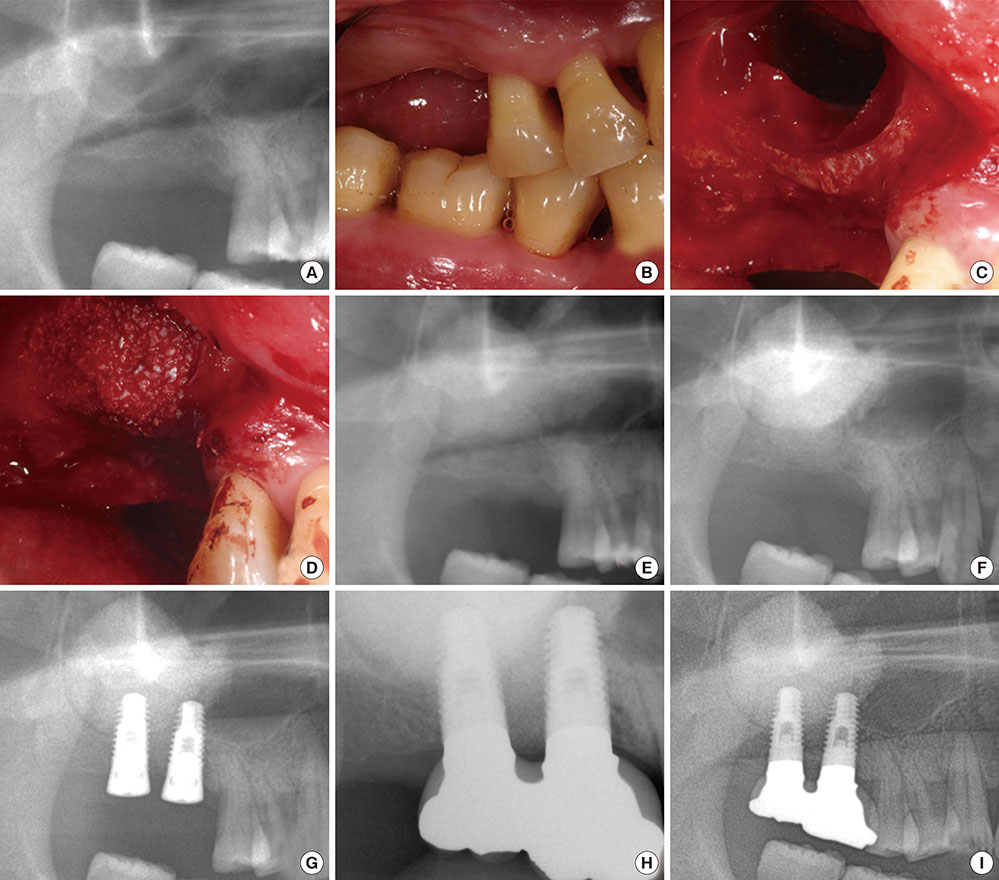
Figure 3 Clinical and radiographic images of patient 3. (A) Panoramic view before the maxillary sinus graft procedure. The right molars were missing. (B) Clinical view on the buccal side of the edentulous area. (C) Window opening of the lateral wall of the right sinus. (D) Demineralized bovine bone material grafting through the window opening. (E) Panoramic view after the maxillary sinus graft procedure. (F) Panoramic view at 6 months after the maxillary sinus graft procedure. Radiopacity was increased in the graft area. (G) Panoramic view after the fixture installation. (H) The final restoration was delivered 13 months after the maxillary sinus graft procedure. (I) Four years and 4 months after the maxillary sinus graft procedure.
Reference
-
1. Kim YS, Kim SH, Kim KH, Jhin MJ, Kim WK, Lee YK, et al. Rabbit maxillary sinus augmentation model with simultaneous implant placement: differential responses to the graft materials. J Periodontal Implant Sci. 2012; 42:204–211.
Article2. Peng W, Kim IK, Cho HY, Pae SP, Jung BS, Cho HW, et al. Assessment of the autogenous bone graft for sinus elevation. J Korean Assoc Oral Maxillofac Surg. 2013; 39:274–282.
Article3. Hiatt WH, Schallhorn RG, Aaronian AJ. The induction of new bone and cementum formation. IV. Microscopic examination of the periodontium following human bone and marrow allograft, autograft and nongraft periodontal regenerative procedures. J Periodontol. 1978; 49:495–512.
Article4. Browaeys H, Bouvry P, De Bruyn H. A literature review on biomaterials in sinus augmentation procedures. Clin Implant Dent Relat Res. 2007; 9:166–177.
Article5. Schlegel KA, Fichtner G, Schultze-Mosgau S, Wiltfang J. Histologic findings in sinus augmentation with autogenous bone chips versus a bovine bone substitute. Int J Oral Maxillofac Implants. 2003; 18:53–58.6. Park HN, Han SH, Kim KH, Lee SC, Park YJ, Lee SH, et al. A study on the safety and efficacy of bovine bone-derived bone graft material (OCS-B). J Korean Acad Periodontol. 2005; 35:335–343.
Article7. Yeo SI, Park SH, Noh WC, Park JW, Lee JM, Suh JY. A comparative analysis of basic characteristics of several deproteinized bovine bone substitutes. J Korean Acad Periodontol. 2009; 39:149–156.
Article8. Tatum H Jr. Maxillary and sinus implant reconstructions. Dent Clin North Am. 1986; 30:207–229.9. Boyne PJ, James RA. Grafting of the maxillary sinus floor with autogenous marrow and bone. J Oral Surg. 1980; 38:613–616.10. Kim YK, Cho YS, Yun PY. Assessment of dentists' subjective satisfaction with a newly developed device for maxillary sinus membrane elevation by the crestal approach. J Periodontal Implant Sci. 2013; 43:308–314.
Article11. Lee YM, Shin SY, Kim JY, Kye SB, Ku Y, Rhyu IC. Bone reaction to bovine hydroxyapatite for maxillary sinus floor augmentation: histologic results in humans. Int J Periodontics Restorative Dent. 2006; 26:471–481.12. Son WK, Shin SY, Yang SM, Kye SB. Maxillary sinus floor augmentation with anorganic bovine bone: Histologic evaluation in humans. J Korean Acad Periodontol. 2009; 39:95–102.
Article13. Hallman M, Sennerby L, Lundgren S. A clinical and histologic evaluation of implant integration in the posterior maxilla after sinus floor augmentation with autogenous bone, bovine hydroxyapatite, or a 20:80 mixture. Int J Oral Maxillofac Implants. 2002; 17:635–643.14. Maiorana C, Sigurta D, Mirandola A, Garlini G, Santoro F. Sinus elevation with alloplasts or xenogenic materials and implants: an up-to-4-year clinical and radiologic follow-up. Int J Oral Maxillofac Implants. 2006; 21:426–432.15. Sohn DS, Lee JS, Ahn MR, Shin HI. New bone formation in the maxillary sinus without bone grafts. Implant Dent. 2008; 17:321–331.
Article16. Artzi Z, Tal H, Dayan D. Porous bovine bone mineral in healing of human extraction sockets. Part 1: histomorphometric evaluations at 9 months. J Periodontol. 2000; 71:1015–1023.
Article17. Lee DW, Pi SH, Lee SK, Kim EC. Comparative histomorphometric analysis of extraction sockets healing implanted with bovine xenografts, irradiated cancellous allografts, and solvent-dehydrated allografts in humans. Int J Oral Maxillofac Implants. 2009; 24:609–615.18. Valentini P, Abensur DJ. Maxillary sinus grafting with anorganic bovine bone: a clinical report of long-term results. Int J Oral Maxillofac Implants. 2003; 18:556–560.19. Byun YK, Park JB, Kim TI, Seol YJ, Lee YM, Ku Y, et al. Bone reaction to bovine hydroxyapatite grafted in the mandibular defects of beagle dogs. J Korean Acad Periodontol. 2006; 36:39–49.
Article20. Park JB, Lee JY, Park HN, Seol YJ, Park YJ, Rhee SH, et al. Osteopromotion with synthetic oligopeptide-coated bovine bone mineral in vivo. J Periodontol. 2007; 78:157–163.
Article21. Park JB, Lee JY, Park YJ, Rhee SH, Lee SC, Kim TI, et al. Enhanced bone regeneration in beagle dogs with bovine bone mineral coated with a synthetic oligopeptide. J Periodontol. 2007; 78:2150–2155.
Article22. Hieu PD, Chung JH, Yim SB, Hong KS. A radiographical study on the changes in height of grafting materials after sinus lift: a comparison between two types of xenogenic materials. J Periodontal Implant Sci. 2010; 40:25–32.
Article
- Full Text Links
-
- Actions
-
Cited
- CITED
-
- Close
- Share
-
- Similar articles
-
- The literature review on the sinus bone graft using deproteinized bovine bone mineral with lateral approach
- Maxillary sinus bone graft using particulated ramal autobone and bovine bone
- Clinical Evaluation of Simultaneous Implants Placement Following Augmentation of the Maxillary Sinus with Deproteinized Bovine Bone
- The Efficacy of the Graft Materials after Sinus Elevation: Retrospective Comparative Study Using Panoramic Radiography
- Evaluation of Linear and Volumetric Changes in Bone Grafts after Sinus Floor Elevation using a Lateral Approach
