Iatrogenic Intradural Lumbosacral Cyst Following Epiduroscopy
- Affiliations
-
- 1Department of Neurosurgery, Seoul St. Mary's Hospital, The Catholic University College of Medicine, Seoul, Korea. ckpmd@catholic.ac.kr
- KMID: 2018259
- DOI: http://doi.org/10.3340/jkns.2012.52.5.491
Abstract
- We report a rare complication of iatrogenic spinal intradural following minimally invasive extradural endoscopic procedues in the lumbo-sacral spines. To our knowledge, intradural cyst following epiduroscopy has not been reported in the literature. A 65-year-old woman with back pain related with previous lumbar disc surgery underwent endoscopic epidural neuroplasty and nerve block, but her back pain much aggravated after this procedure. Postoperative magnetic resonance imaging revealed a large intradural cyst from S1-2 to L2-3 displacing the nerve roots anteriorly. On T1 and T2-weighted image, the signal within the cyst had the same intensity as cerebrospinal fluid. The patient underwent partial laminectomy of L5 and intradural exploration, and fenestration of the cystic wall was accomplished. During operation, the communication between the cyst and subarachnoid space was not identified, and the content of the cyst was the same as that of cerebrospinal fluid. Postoperatively, the pain attenuated immediately. Incidental durotomy which occurred during advancing the endoscope through epidural space may be the cause of formation of the intradural cyst. Intrdural cyst should be considered, if a patient complains of new symptoms such as aggravation of back pain after epiduroscopy. Surgical treatment, simple fenestration of the cyst may lead to improved outcome. All the procedures using epiduroscopy should be performed with caution.
Keyword
MeSH Terms
Figure
-
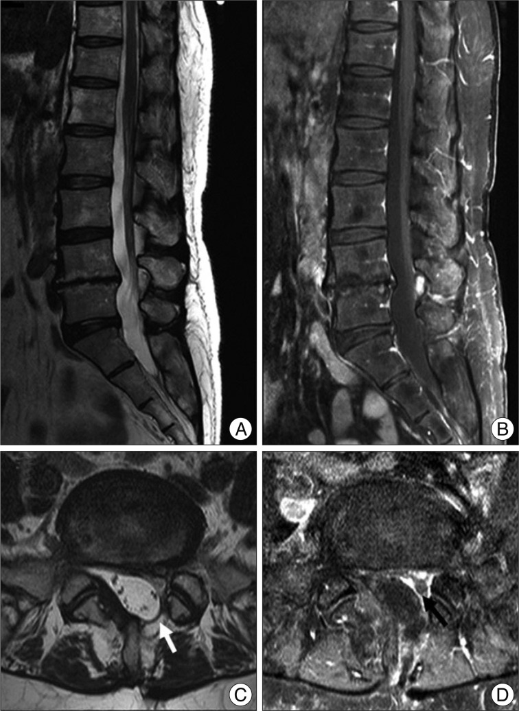
Fig. 1 Lumbar sagittal T2 weighted (A), Gd-enhanced T1 weighted (B), axial T2 weighted (C), and Gd-enhanced T1 weighted (D) magnetic resonance imaging scans showing the postoperative changes of L4-5 level, which presenting no recurrence of disc herniation except for the findings of mild adhesion and left partial hemilaminectomy of L4 (white and black arrows).

Fig. 2 A fluoroscopic view showing the endoscopic epidural neuroplasty. An epidural catheter is inserted into the epidural space of L5-S1 levels through the sacral hiatus. The tip of epidural catheter is located just around right L5 root.
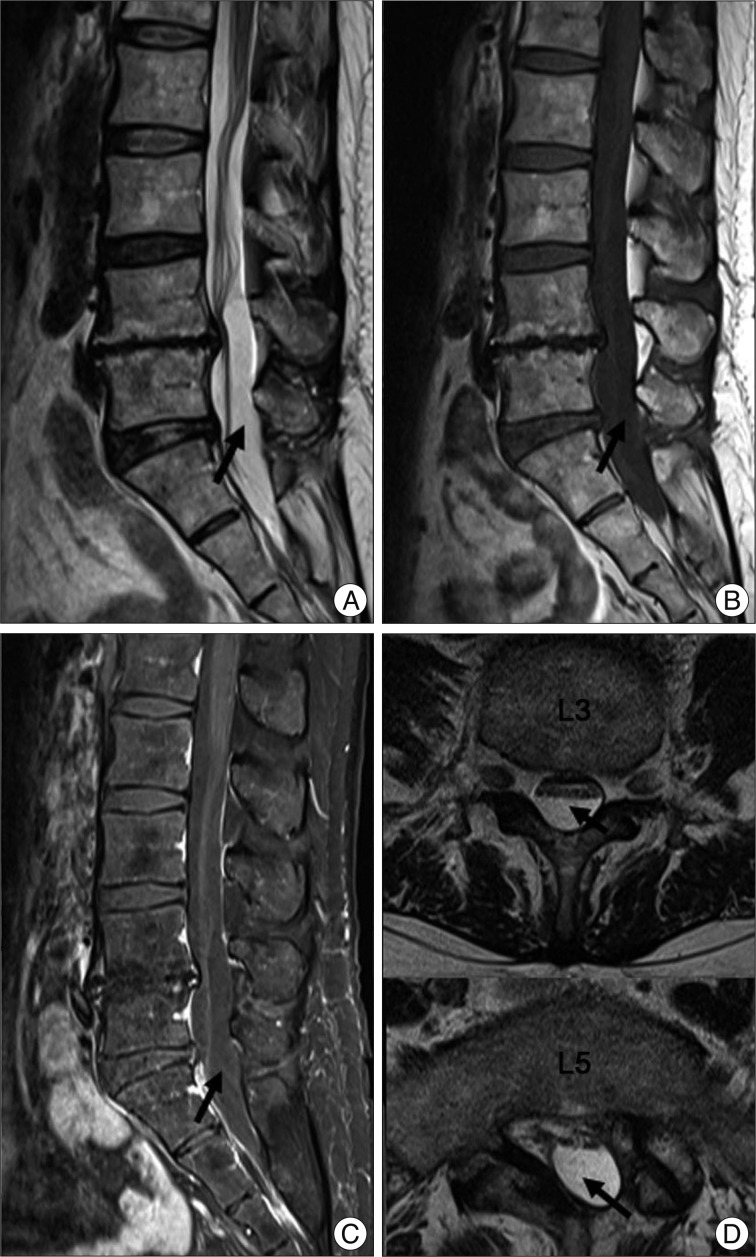
Fig. 3 Sagittal T2 (A), T1 (B), Gd-enhanced T1 (C) and axial T2 (D) weighted magnetic resonance imaging scans showing an intradural cystic lesion (black arrows), spanning from L2-3 to S2-3 level and displacing cauda equina anteriorly. The cyst fluid having the same signal intensity as cerebrospinal fluid.

Fig. 4 Myelography-computed tomography images showing an intradural cyst spanning from L4-5 to S2-3 level (black arrow) (A). The contrast media is partially filled up in the cystic cavity of the lower spines (black arrows) (B).
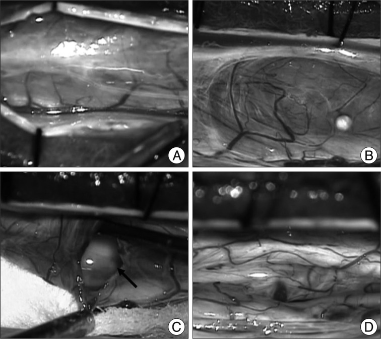
Fig. 5 Intraoperative photographs. A : After hemilaminectomy, the dura mater is opened. B : A large arachnoid cyst, which presents well demarcated by thin and transparent wall and contains cerebrospinal fluid, is observed in the dorsal part of the intradural space, compressing and displacing the cauda equina anteriorly. C : Cystic wall and enlargement of the window were performed. No connection of the cyst with subarachnoid space is noticed. D : After partial removal of cyst wall, nerve roots are exposed and decompressed.
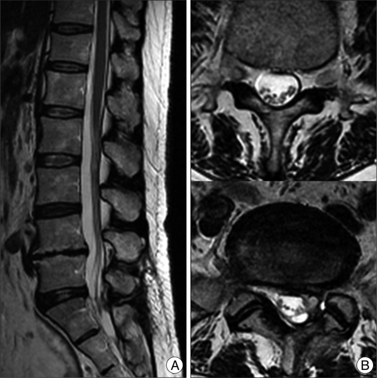
Fig. 6 Sagittal (A) and axial (B) postoperative magnetic resonance imaging scans showing complete disappearance of the intradural cyst.
Cited by 3 articles
-
The outcome of epiduroscopy treatment in patients with chronic low back pain and radicular pain, operated or non-operated for lumbar disc herniation: a retrospective study in 88 patients
Derya Burcu Hazer, Arsal Acarbaş, Hans Eric Rosberg
Korean J Pain. 2018;31(2):109-115. doi: 10.3344/kjp.2018.31.2.109.Complication of epiduroscopy: a brief review and case report
Maurizio Marchesini, Edoardo Flaviano, Valentina Bellini, Marco Baciarello, Elena Giovanna Bignami
Korean J Pain. 2018;31(4):296-304. doi: 10.3344/kjp.2018.31.4.296.Unintended Complication of Intracranial Subdural Hematoma after Percutaneous Epidural Neuroplasty
Sung Bum Kim, Min Ki Kim, Kee D. Kim, Young Jin Lim
J Korean Neurosurg Soc. 2014;55(3):170-172. doi: 10.3340/jkns.2014.55.3.170.
Reference
-
1. Avellanal M, Diaz-Reganon G. Interlaminar approach for epiduroscopy in patients with failed back surgery syndrome. Br J Anaesth. 2008; 101:244–249. PMID: 18552347.
Article2. Gillespie G, MacKenzie P. Epiduroscopy--a review. Scott Med J. 2004; 49:79–81. PMID: 15462218.
Article3. Igarashi T, Hirabayashi Y, Seo N, Saitoh K, Fukuda H, Suzuki H. Lysis of adhesions and epidural injection of steroid/local anaesthetic during epiduroscopy potentially alleviate low back and leg pain in elderly patients with lumbar spinal stenosis. Br J Anaesth. 2004; 93:181–187. PMID: 15194631.
Article4. Kriss TC, Kriss VM. Symptomatic spinal intradural arachnoid cyst development after lumbar myelography. Case report and review of the literature. Spine (Phila Pa 1976). 1997; 22:568–572. PMID: 9076891.
Article5. Mao HQ, Yang HL, Geng DC, Bao ZH, Tang TS. Spinal extradural arachnoid cyst following percutaneous vertebroplasty. Eur Spine J. 2011; 20(Suppl 2):S206–S210. PMID: 20835874.
Article6. Nabors MW, Pait TG, Byrd EB, Karim NO, Davis DO, Kobrine AI, et al. Updated assessment and current classification of spinal meningeal cysts. J Neurosurg. 1988; 68:366–377. PMID: 3343608.
Article7. Nottmeier EW, Wharen RE, Patel NP. Iatrogenic intradural spinal arachnoid cyst as a complication of lumbar spine surgery. J Neurosurg Spine. 2009; 11:344–346. PMID: 19769517.
Article
- Full Text Links
-
- Actions
-
Cited
- CITED
-
- Close
- Share
-
- Similar articles
-
- Extensive Intradural Epidermoid Cysts with Cauda Equina Syndrome in the Lumbosacral Spine: Case Report
- Multiple Lumbar Intradural Dermoid Cysts without Spinal Dysraphism
- Intradural Epidermoid Cyst: A Case Report
- Gas-Filled Intradural Cyst within the Cauda Equine
- A Rare Case of Thoracic Intradural Epidermoid Cyst after Spinal Cord Stimulator Insertion: A Case Report
