Characteristics of bony changes and tooth displacement in the mandibular cystic lesion involving the impacted third molar
- Affiliations
-
- 1Department of Oral and Maxillofacial Surgery, Gangnam Severance Hospital, Yonsei University College of Dentistry, Seoul, Korea. omshuh@yuhs.ac
- 2Department of Oral and Maxillofacial Surgery, Yongin Severance Hospital, Yonsei University College of Dentistry, Seoul, Korea.
- 3Department of Oral and Maxillofacial Radiology, Yongin Severance Hospital, Yonsei University College of Dentistry, Seoul, Korea.
- KMID: 2005452
- DOI: http://doi.org/10.5125/jkaoms.2014.40.5.225
Abstract
OBJECTIVES
The purpose of this retrospective study is to find the differentiating characteristics of cystic and cystic-appearing lesions that involve the impacted mandibular third molar by analyzing panoramic radiographs and computed tomography images, and to aid the preoperative diagnosis.
MATERIALS AND METHODS
Eighty-one patients who had a mandibular cystic or cystic-appearing lesion that involved impacted mandibular third molar and underwent cyst enucleation were included in the study. The preoperative panoramic radiograph and computed tomography findings were analyzed in accordance to the histopathologic type.
RESULTS
Most of the cystic lesions containing the mandibular third molar were diagnosed as a dentigerous cyst (77.8%). The occurrence of mesio-distal displacement of the third molar was more frequent in the odontogenic keratocyst (71.4%) and in the ameloblastoma (85.7%) than in the dentigerous cyst (19.1%). Downward displacement was primarily observed in each group. Odontogenic keratocyst and ameloblastoma showed more aggressive growth pattern with higher rate of bony discontinuity and cortical bone expansion than in dentigerous cyst.
CONCLUSION
When evaluating mandibular cystic lesions involving the impacted mandibular third molar, dentigerous cyst should first be suspected. However, when the third molar displacement and cortical bone absorption are observed, then odontogenic keratocyst or ameloblastoma should be considered.
MeSH Terms
Figure
-
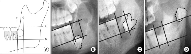
Fig. 1 Tooth displacement patterns on panoramic radiographs. Any position that did not deviate from the described standard was considered normal. A. a: Occlusal plane of the mesial teeth. b: The line parallel to (a), extending to the root tips of the second molar. c: The line perpendicular to (a) and tangent to the height of the distal contour of the second molar. d: The line parallel to (c) and located behind the length of the mesio-buccal width of the second molar. B. Downward displacement. C. Backward displacement. D. Back-upward displacement.
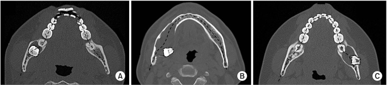
Fig. 2 Tooth displacement patterns on computed tomography imaging. A. No displacement. B. Lingual displacement. C. Buccal displacement.
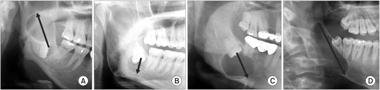
Fig. 3 Growth patterns of the lesion on displayed panoramic radiographs. A. Back-upward (arrow). B. Downward (arrow). C. Down-forward (arrow). D. Down-forward and back-upward (arrow).

Fig. 4 Growth patterns of the lesion shown on computed tomography imaging. A. Buccal (asterisk). B. Bucco-lingual. C. Lingual (asterisk).
Reference
-
1. Devenney-Cakir B, Subramaniam RM, Reddy SM, Imsande H, Gohel A, Sakai O. Cystic and cystic-appearing lesions of the mandible: review. AJR Am J Roentgenol. 2011; 196:WS66–WS77. PMID: 21606244.2. Knutsson K, Brehmer B, Lysell L, Rohlin M. Pathoses associated with mandibular third molars subjected to removal. Oral Surg Oral Med Oral Pathol Oral Radiol Endod. 1996; 82:10–17. PMID: 8843448.
Article3. Scholl RJ, Kellett HM, Neumann DP, Lurie AG. Cysts and cystic lesions of the mandible: clinical and radiologic-histopathologic review. Radiographics. 1999; 19:1107–1124. PMID: 10489168.
Article4. Dunfee BL, Sakai O, Pistey R, Gohel A. Radiologic and pathologic characteristics of benign and malignant lesions of the mandible. Radiographics. 2006; 26:1751–1768. PMID: 17102048.
Article5. Yoshiura K, Higuchi Y, Araki K, Shinohara M, Kawazu T, Yuasa K, et al. Morphologic analysis of odontogenic cysts with computed tomography. Oral Surg Oral Med Oral Pathol Oral Radiol Endod. 1997; 83:712–718. PMID: 9195629.
Article6. Eun SA, Kim KD, Park CS. Differential diagnosis between odontogenic keratocyst and ameloblastoma by computed tomography. Korean J Oral Maxillofac Radiol. 2002; 32:89–97.7. Tsukamoto G, Sasaki A, Akiyama T, Ishikawa T, Kishimoto K, Nishiyama A, et al. A radiologic analysis of dentigerous cysts and odontogenic keratocysts associated with a mandibular third molar. Oral Surg Oral Med Oral Pathol Oral Radiol Endod. 2001; 91:743–747. PMID: 11402292.
Article8. Saravana GH, Subhashraj K. Cystic changes in dental follicle associated with radiographically normal impacted mandibular third molar. Br J Oral Maxillofac Surg. 2008; 46:552–553. PMID: 18406023.
Article9. Baykul T, Saglam AA, Aydin U, Başak K. Incidence of cystic changes in radiographically normal impacted lower third molar follicles. Oral Surg Oral Med Oral Pathol Oral Radiol Endod. 2005; 99:542–545. PMID: 15829874.
Article10. Cankurtaran CZ, Branstetter BF 4th, Chiosea SI, Barnes EL Jr. Best cases from the AFIP: ameloblastoma and dentigerous cyst associated with impacted mandibular third molar tooth. Radiographics. 2010; 30:1415–1420. PMID: 20833858.11. Haring JI, Van Dis ML. Odontogenic keratocysts: a clinical, radiographic, and histopathologic study. Oral Surg Oral Med Oral Pathol. 1988; 66:145–153. PMID: 2457195.12. Güven O, Keskin A, Akal UK. The incidence of cysts and tumors around impacted third molars. Int J Oral Maxillofac Surg. 2000; 29:131–135. PMID: 10833151.
Article13. Reichart PA, Philipsen HP, Sonner S. Ameloblastoma: biological profile of 3677 cases. Eur J Cancer B Oral Oncol. 1995; 31B:86–99. PMID: 7633291.
Article14. Theodorou SJ, Theodorou DJ, Sartoris DJ. Imaging characteristics of neoplasms and other lesions of the jawbones: part 1. Odontogenic tumors and tumorlike lesions. Clin Imaging. 2007; 31:114–119. PMID: 17320778.15. Brannon RB. The odontogenic keratocyst. A clinicopathologic study of 312 cases. Part I. Clinical features. Oral Surg Oral Med Oral Pathol. 1976; 42:54–72. PMID: 1065842.16. Werkmeister R, Fillies T, Joos U, Smolka K. Relationship between lower wisdom tooth position and cyst development, deep abscess formation and mandibular angle fracture. J Craniomaxillofac Surg. 2005; 33:164–168. PMID: 15878516.
Article17. Soh BC, Heo MS, An CH, Choi M, Lee SS, Choi SC, et al. Radiographic differential diagnosis between ameloblastoma and odontogenic keratocyst: with emphasis on CT. Korean J Oral Maxillofac Radiol. 2002; 32:167–173.
- Full Text Links
-
- Actions
-
Cited
- CITED
-
- Close
- Share
-
- Similar articles
-
- Migration of mandibular third molar to the condyle without cystic change: a case report
- Cone beam computed tomography findings of ectopic mandibular third molar in the mandibular condyle: report of a case
- Eruption Guidance of Horizontally Impacted Permanent First Molar with Primary Retention of Primary Second Molars: Case Reports
- Pressure Root Resorption of the Second Molar Caused by Third Molar Impaction: A Case Report of Severely Resorbed Root with Vital Pulp
- Surgical-orthodontic treatment of impacted teeth displaced by unicystic ameloblastoma
