An Unusual Radiologic Manifestation of Pulmonary Tuberculosis with Bilateral Multiple Lung Nodules and Diffuse Alveolar Hemorrhage: A Case Report
- Affiliations
-
- 1Department of Radiology, Chonnam National University Hospital, Gwangju, Korea. sunaura@hanmail.net
- 2Department of Radiology, Chonnam National University Hwasun Hospital, Hwasun, Korea.
- KMID: 2002926
- DOI: http://doi.org/10.3348/jksr.2011.65.6.585
Abstract
- Pulmonary tuberculosis presenting as bilateral multiple lung nodules or diffuse alveolar hemorrhage is very rare. Here, we report a case of pulmonary tuberculosis presenting as bilateral multiple lung nodules and diffuse alveolar hemorrhage mimicking granulomatous vasculitis, such as Wegener's granulomatosis.
MeSH Terms
Figure
-
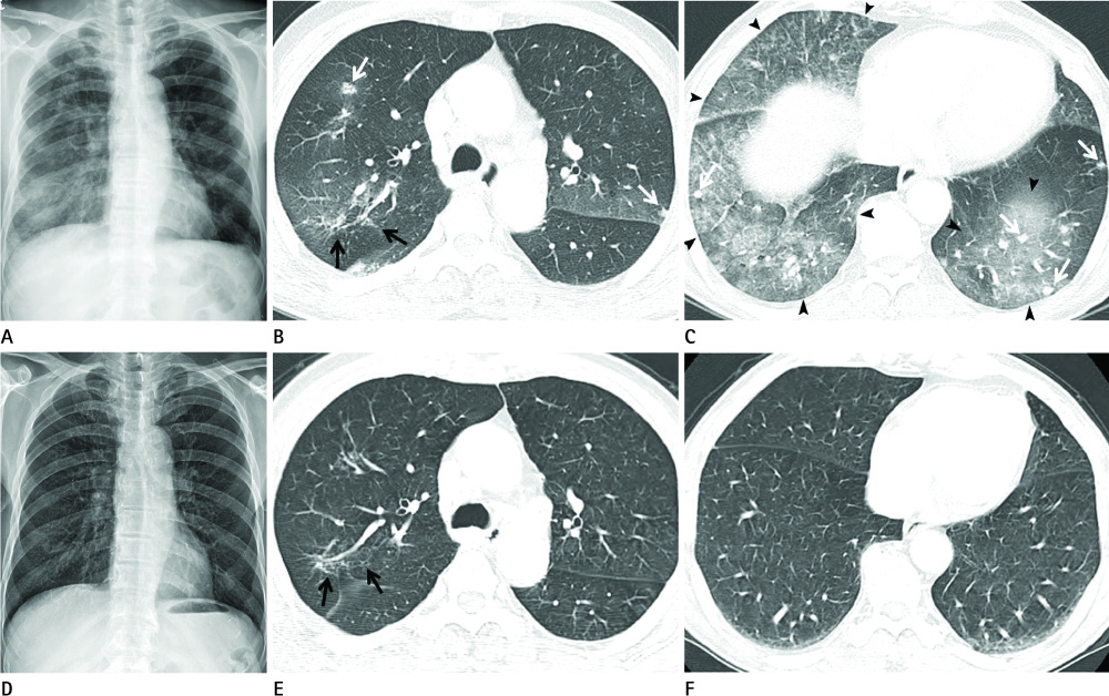
Fig. 1 A 70-year-old man with pulmonary tuberculosis. A. Initial chest PA radiogram shows multiple nodular opacities with mildly and diffusely increased opacity in both lungs and consolidation in right lower lung. B, C. Initial chest CT scan shows multiple bilateral nodules with ground glass attenuation halo (white arrows in B, C) mixed with crazy-paving pattern lesions (arrowheads in C) in both lung fields with more prominent involvement in the both lower lobes. Focal area of fibrotic old inflammatory lesion suggesting inactive pulmonary tuberculosis is also shown in posterior segment of right upper lobe (black arrows in B). D. Follow up chest PA radiogram after anti-tuberculosis medication shows improvement of multiple nodular opacities in both lungs and consolidation in right lower lung. E, F. Follow up chest CT after anti-tuberculosis medication shows complete improvement of multiple bilateral nodules and crazy-paving pattern lesions in both lungs with remained focal area of fibrotic old inflammatory lesion in posterior segment of RUL (black arrows in E). Note.-PA = posteroanterior
Reference
-
1. Leung AN. Pulmonary tuberculosis: the essentials. Radiology. 1999; 210:307–322.2. Fabreguet I, Francis F, Lemery M, Choudat L, Papo T, Sacre K. A 76-year-old man with multiple pulmonary nodules. Chest. 2009; 135:1094–1097.3. Yoshitomi A, Ono T, Sato A, Nakamura H, Chida K. [Pulmonary tuberculosis with diabetes mellitus, presenting multiple nodular shadows]. Kansenshogaku Zasshi. 1998; 72:561–563.4. Keung YK, Nugent K, Jumper C, Cobos E. Mycobacterium tuberculosis infection masquerading as diffuse alveolar hemorrhage after autologous stem cell transplant. Bone Marrow Transplant. 1999; 23:737–738.5. Marruchella A, Corpolongo A, Tommasi C, Lauria FN, Narciso P. A case of pulmonary tuberculosis presenting as diffuse alveolar haemorrhage: is there a role for anticardiolipin antibodies? BMC Infect Dis. 2010; 10:33.6. Espinosa G, Cervera R, Font J, Asherson RA. The lung in the antiphospholipid syndrome. Ann Rheum Dis. 2002; 61:195–198.7. Yeh JJ, Chen SC, Teng WB, Chou CH, Hsieh SP, Lee TL, et al. Identifying the most infectious lesions in pulmonary tuberculosis by high-resolution multi-detector computed tomography. Eur Radiol. 2010; 20:2135–2145.8. Pinto PS. The CT Halo Sign. Radiology. 2004; 230:109–110.9. Elkayam O, Caspi D, Lidgi M, Segal R. Auto-antibody profiles in patients with active pulmonary tuberculosis. Int J Tuberc Lung Dis. 2007; 11:306–310.
- Full Text Links
-
- Actions
-
Cited
- CITED
-
- Close
- Share
-
- Similar articles
-
- Sarcoidosis Presenting with Multiple Lung Parenchymal Nodules
- Diffuse Large B-Cell Lymphoma Manifesting as Miliary Nodules in the Lung: A Case Report
- A case of pulmonary diffuse alveolar amyloidosis localized in the lung
- A Case of Pulmonary Tuberculosis Presenting as Diffuse Interstitial Lung Disease Associated with the Lymphadenopathy of Mediastinum and Abdomen
- Alveolar Septal Pulmonary Amyloidosis: A Case Report
