J Korean Soc Magn Reson Med.
2014 Sep;18(3):219-224. 10.13104/jksmrm.2014.18.3.219.
Pseudoradial Tear of the Medial Meniscus: A Relatively Common Potential Pitfall
- Affiliations
-
- 1Department of Radiology, Hallym University Dongtan Sacred Heart Hospital, Hwaseong, Korea. jachoi88@gmail.com
- 2Department of Radiology, Seoul National University Bundang Hospital, Seongnam, Korea.
- KMID: 1999921
- DOI: http://doi.org/10.13104/jksmrm.2014.18.3.219
Abstract
- PURPOSE
To determine the incidence of truncated triangle appearance of anterior horn (AH) to body of medial meniscus (MM) and determine its clinical significance.
MATERIALS AND METHODS
IRB approval was obtained, and informed consent waived for this study. The criteria of "pseudoradial tear" was truncated triangle appearance of the tip of AH to body of MM on one or more coronal images with adjacent fluid signal intensity at the blunted tip. Two musculoskeletal radiologists retrospectively evaluated 485 knee MR images independently for the presence and number of sections with "pseudoradial tear" of AH to body of MM using proton density-weighted coronal MR images. Inter-and intraobserver agreement was calculated using kappa coefficients. Medical records were reviewed for arthroscopic correlation.
RESULTS
A pseudoradial tear in the AH to body of MM was present in 381 (78.6%) patients. Locations were 112 in AH (29.4%), 143 in AH to body (37.5%), and 126 in body (33.1%). Number of consecutive sections of pseudoradial tear were 1 in 100 (26.2%), 2 in 164 (43.0%), 3 in 94 (24.7%), 4 in 21 (5.5%), and 5 in 2 (0.5%). Interobserver agreement was 0.99 for presence and 0.43 for number of sections of pseudoradial tear. Arthroscopies were performed in 96 patients and none of the pseudoradial tears were proven as true radial tears on arthroscopy.
CONCLUSION
Pseudoradial tears are frequently seen in AH to body of MM on coronal MR images and may be another pitfall that a radiologist needs to be aware of and be able to differentiate from true radial tear.
Keyword
MeSH Terms
Figure
-
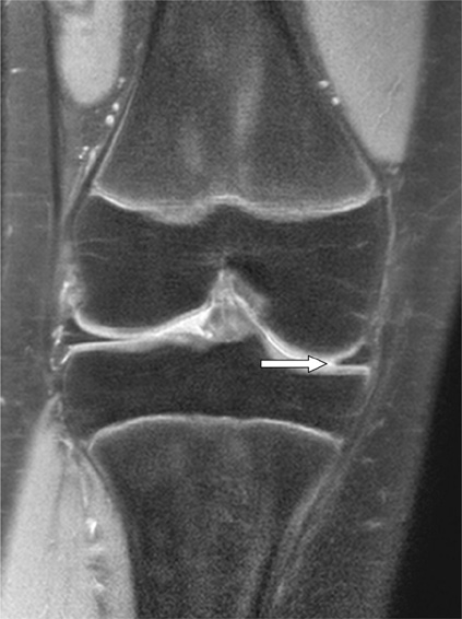
Fig. 1 Pseudoradial tear at the junction of anterior horn to body of the medial meniscus in a 11-year-old girl on proton densityweighted spectral presaturation inversion recovery (SPIR) coronal image (TR/TE, 2718/7.617; 3-mm section thickness) shown as blunting of the tip of the medial meniscus (arrow). Meniscus was intact at arthroscopy.

Fig. 2 Pseudoradial tear at the junction of anterior horn to body of the medial meniscus in a 39-year-old women on proton density-weighted coronal image (TR/TE, 2651/12; 4-mm section thickness) shows adjacent fluid signal intensity at the blunted tip of the medial meniscus (arrow). Meniscus was intact at arthroscopy.
Reference
-
1. Magee T, Shapiro M, Williams D. MR accuracy and arthroscopic incidence of meniscal radial tears. Skeletal Radiol. 2002; 31:686–689.2. Justice WW, Quinn SF. Error patterns in the MR imaging evaluation of menisci of the knee. Radiology. 1995; 196:617–621.3. De Smet AA, Tuite MJ, Norris MA, Swan JS. MR diagnosis of meniscal tears: analysis of causes of errors. AJR Am J Roentgenol. 1994; 163:1419–1423.4. De Smet AA, Nathan DH, Graf BK, Haaland BA, Fine JP. Clinical and MRI findings assciated with false positive knee MR diagnoses of medial meniscal tears. AJR Am J Roentgenol. 2008; 191:93–99.5. De Smet A, Graf B. Meniscal tears missed on MR imaging: relationship to meniscal tear patterns and anterior cruciate ligament tears. AJR Am J Roentgenol. 1994; 162:905–911.6. Watanabe AT, Carter BC, Teitelbaum GP, Seeger LL, Bradley WG. Normal variations in MR imaging of the knee: appearance and frequency. AJR Am J Roentgenol. 1989; 153:341–344.7. Vahey TN, Bennett HT, Arrington LE, Sholboumo KD, Ng J. MR imaging of the knee: pseudotear of the lateral meniscus caused by the meniscofemoral ligament. AJR Am J Roentgenol. 1990; 154:1237–1239.8. Herman LJ, Beltran J. Pitfalls in MR Imaging of the knee. Radiology. 1988; 167:775–781.9. Pusey E, Lufkin RB, Brown RK, et al. Magnetic resonance imaging artifacts: mechanism and clinical significance. Radiographics. 1986; 6:891–911.10. Firooznia H, Golimbu C, Rafi M. MR imaging of the menisci. Fundamentals of anatomy and pathology. Magn Reson Imaging Clin N Am. 1994; 2:325–347.11. Jee WH, McCauley TR, Kim JM, et al. Meniscal tear configurations: categorization with MR imaging. AJR Am J Roentgenol. 2003; 180:93–97.12. Tuckman GA, Miller WJ, Remo JW, Fritts HM, Rozansky MI. Radial tears of the menisci: MR findings. AJR Am J Roentgenol. 1994; 163:395–400.13. Harper KW, Helms CA, Lambert HS 3rd, Higgins LD. Radial meniscal tears: significance, incidence, and MR appearance. AJR Am J Roentgenol. 2005; 185:1429–1434.14. Messner K, Gao J. The menisci of the knee joint: anatomical and functional characteristics, and a rationale for clinical treatment. J Anat. 1998; 193:161–178.15. Magee T, Williams D. Detection of meniscal tears and marrow lesions using coronal MRI. AJR Am J Roentgenol. 2004; 183:1469–1473.
- Full Text Links
-
- Actions
-
Cited
- CITED
-
- Close
- Share
-
- Similar articles
-
- Non-Operative Treatment of the Degenerative Medial Meniscus Posterior Root Tear
- Medial Meniscus Posterior Horn Root Tear in Adolescent during Sport Activity: A Case Report
- Avulsion Fracture of the Anterior Medial Meniscus Root
- Clinical Characteristics of Isolated Meniscal Tear
- Clinical Study of Meniscus Tears in Koreans
