Restor Dent Endod.
2013 Feb;38(1):26-30. 10.5395/rde.2013.38.1.26.
Apical foramen morphology according to the length of merged canal at the apex
- Affiliations
-
- 1Department of Conservative Dentistry, Chosun University School of Dentistry, Gwangju, Korea. rootcanal@hanmail.net
- KMID: 1995473
- DOI: http://doi.org/10.5395/rde.2013.38.1.26
Abstract
OBJECTIVES
The aim of this study was to investigate the relationship between the apical foramen morphology and the length of merged canal at the apex in type II root canal system.
MATERIALS AND METHODS
This study included intact extracted maxillary and mandibular human premolars (n = 20) with fully formed roots without any visible signs of external resorption. The root segments were obtained by removing the crown 1 mm beneath the cementum-enamel junction (CEJ) using a rotary diamond disk. The distance between the file tip and merged point of joining two canals was defined as Lj. The roots were carefully sectioned at 1 mm from the apex by a slow-speed water-cooled diamond saw. All cross sections were examined under the microscope at x50 magnification and photographed to estimate the shape of the apical foramen. The longest and the shortest diameter of apical foramen was measured using ImageJ program (1.44p, National Institutes of Health). Correlation coefficient was calculated to identify the link between Lj and the apical foramen shape by Pearson's correlation.
RESULTS
The average value of Lj was 3.74 mm. The average of proportion (P), estimated by dividing the longest diameter into the shortest diameter of the apical foramen, was 3.64. This study showed a significant negative correlation between P and Lj (p < 0.05).
CONCLUSIONS
As Lj gets longer, the apical foramen becomes more ovally shaped. Likewise, as it gets shorter, the apical foramen becomes more flat shaped.
Keyword
MeSH Terms
Figure
-

Figure 1 The distance between the file tip and merging point of two joining canals (Lj) on radiograph.
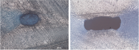
Figure 2 The apical foramen shape (×50).
Cited by 1 articles
-
Surgical endodontic management of infected lateral canals of maxillary incisors
Ji-Hyun Jang, Jung-Min Lee, Jin-Kyu Yi, Sung-Baik Choi, Sang-Hyuk Park
Restor Dent Endod. 2015;40(1):79-84. doi: 10.5395/rde.2015.40.1.79.
Reference
-
1. Baratto Filho F, Zaitter S, Haragushiku GA, de Campos EA, Abuabara A, Correr GM. Analysis of the internal anatomy of maxillary first molars by using different methods. J Endod. 2009. 35:337–342.
Article2. Olson DG, Roberts S, Joyce AP, Collins DE, McPherson JC 3rd. Unevenness of the apical constriction in human maxillary central incisors. J Endod. 2008. 34:157–159.
Article3. Hassanien EE, Hashem A, Chalfin H. Histomorphometric study of the root apex of mandibular premolar teeth: an attempt to correlate working length measured with electronic and radiograph methods to various anatomic positions in the apical portion of the canal. J Endod. 2008. 34:408–412.
Article4. Vertucci FJ. Root canal anatomy of the human permanent teeth. Oral Surg Oral Med Oral Pathol. 1984. 58:589–599.
Article5. Olson AK, Goerig AC, Cavataio RE, Luciano J. The ability of the radiograph to determine the location of the apical foramen. Int Endod J. 1991. 24:28–35.
Article6. Jafarzadeh H, Wu YN. The C-shaped root canal configuration: a review. J Endod. 2007. 33:517–523.
Article7. Grande NM, Plotino G, Pecci R, Bedini R, Pameijer CH, Somma F. Micro-computerized tomographic analysis of radicular and canal morphology of premolars with long oval canals. Oral Surg Oral Med Oral Pathol Oral Radiol Endod. 2008. 106:e70–e76.
Article8. Wu MK, Barkis D, Roris A, Wesselink PR. Does the first file to bind correspond to the diameter of the canal in the apical region? Int Endod J. 2002. 35:264–267.
Article9. Wu MK, R'oris A, Barkis D, Wesselink PR. Prevalence and extent of long oval canals in the apical third. Oral Surg Oral Med Oral Pathol Oral Radiol Endod. 2000. 89:739–743.
Article10. Martos J, Lubian C, Silveira LF, Suita de Castro LA, Ferrer Luque CM. Morphologic analysis of the root apex in human teeth. J Endod. 2010. 36:664–667.
Article11. Wu MK, Wesselink PR. A primary observation on the preparation and obturation of oval canals. Int Endod J. 2001. 34:137–141.
Article12. Weiger R, Bartha T, Kalwitzki M, Löst C. A clinical method to determine the optimal apical preparation size. Part I. Oral Surg Oral Med Oral Pathol Oral Radiol Endod. 2006. 102:686–691.
Article13. Jung IY, Seo MA, Fouad AF, Spångberg LS, Lee SJ, Kim HJ, Kum KY. Apical anatomy in mesial and mesiobuccal roots of permanent first molars. J Endod. 2005. 31:364–368.
Article14. Weine FS. Endodontic Therapy. 1996. 5th ed. St Louis: Mosby;243.15. Vertucci F, Seelig A, Gillis R. Root canal morphology of the human maxillary second premolar. Oral Surg Oral Med Oral Pathol. 1974. 38:456–464.
Article16. Jeong EJ, Lee DK, Baek SY, Hwang HK. A comparison of master apical file size according to instrumentation in type II root canal. J Korean Acad Conserv Dent. 2008. 33:435–442.
Article17. Shin JS, Cho YB. Removal patterns of smear layer according to application temparature and time of EDTA. J Korean Acad Conserv Dent. 2002. 27:535–542.
Article18. Lee JK, Park SH, Choi GW. Time-dependent effects of EDTA application on removal of smear layer in the root canal system. J Korean Acad Conserv Dent. 2006. 31:169–178.
Article19. Senia ES. Canal diameter: the forgotten dimension. Dent Today. 2001. 20:58–62.20. Peters OA. Current challenges and concepts in the preparation of root canal systems: a review. J Endod. 2004. 30:559–567.
Article21. Jou YT, Karabucak B, Levin J, Liu D. Endodontic working width: current concepts and techniques. Dent Clin North Am. 2004. 48:323–335.
Article22. Blasković-Subat V, Maricić B, Sutalo J. Asymmetry of the root canal foramen. Int Endod J. 1992. 25:158–164.
Article23. Oyama K, Motoyoshi M, Hirabayashi M, Hosoi K, Shimizu N. Effects of root morphology on stress distribution at the root apex. Eur J Orthod. 2007. 29:113–117.
Article24. Park JW, Lee JK, Ha BH, Choi JH, Perinpanayagam H. Three-dimensional analysis of maxillary first molar mesiobuccal root canal configuration and curvature using micro-computed tomography. Oral Surg Oral Med Oral Pathol Oral Radiol Endod. 2009. 108:437–442.
Article
- Full Text Links
-
- Actions
-
Cited
- CITED
-
- Close
- Share
-
- Similar articles
-
- Morphology of the apical root canal system in Korean mandibular first molar
- Effect of file size in measuring electronic working length of teeth with open apex
- In vivo evaluation of accuracy and consistency of two electronic apex locators
- An evaluation of canal curvature at the apical one third in type II mesial canals of mandibular molars
- An accuracy of the several electronic apex locators on the mesial root canal of the mandibular molar
