Comparison of the centering ability of Wave.One and Reciproc nickel-titanium instruments in simulated curved canals
- Affiliations
-
- 1Department of Conservative Dentistry, Wonkwang University School of Dentistry, Iksan, Korea.
- 2Department of Conservative Dentistry, Pusan National University School of Dentistry, Yangsan, Korea.
- 3Department of Conservative Dentistry, Chonbuk National University School of Dentistry, Jeonju, Korea. endomin@gmail.com
- KMID: 1995472
- DOI: http://doi.org/10.5395/rde.2013.38.1.21
Abstract
OBJECTIVES
The aim of this study was to evaluate the shaping ability of newly marketed single-file instruments, Wave.One (Dentsply-Maillefer) and Reciproc (VDW GmbH), in terms of maintaining the original root canal configuration and curvature, with or without a glide-path.
MATERIALS AND METHODS
According to the instruments used, the blocks were divided into 4 groups (n = 10): Group 1, no glide-path / Wave.One; Group 2, no glide-path / Reciproc; Group 3, #15 K-file / Wave.One; Group 4, #15 K-file / Reciproc. Pre- and post-instrumented images were scanned and the canal deviation was assessed. The cyclic fatigue stress was loaded to examine the cross-sectional shape of the fractured surface. The broken fragments were evaluated under the scanning electron microscope (SEM) for topographic features of the cross-section. Statistically analysis of the data was performed using one-way analysis of variance followed by Tukey's test (alpha = 0.05).
RESULTS
The ability of instruments to remain centered in prepared canals at 1 and 2 mm levels was significantly lower in Group 1 (p < 0.05). The centering ratio at 3, 5, and 7 mm level were not significantly different.
CONCLUSIONS
The Wave.One file should be used following establishment of a glide-path larger than #15.
MeSH Terms
Figure
-
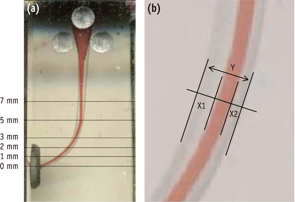
Figure 1 (a) The picture indicates the points at which the canal width was measured after superimposition of pre- and post-operative images; (b) X1 represents the maximum extent of canal movement in one direction and X2 is the movement in the opposite direction. Y is the diameter of the final canal preparation.
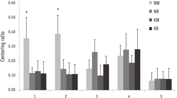
Figure 2 Centering ratio of canals at different apical levels. Values are mean ± SD. *A significant difference was determined at p < 0.05. NW, no glide-path / Wave·One; NR, no glide-path / Reciproc; KW, glidepath with K-file / Wave·One; KR, glidepath with K-file / Reciproc.

Figure 3 Scanning electron micrographs of fracture surface of separated fragments. (a) Reciproc (×200); (b) Wave·One (×180).
Cited by 5 articles
-
Effect of passive ultrasonic agitation during final irrigation on cleaning capacity of hybrid instrumentation
Marcilene Coelho Vinhorte, Eduardo Hideki Suzuki, Maíra Sousa de Carvalho, André Augusto Franco Marques, Emílio Carlos Sponchiado Júnior, Lucas da Fonseca Roberti Garcia
Restor Dent Endod. 2014;39(2):104-108. doi: 10.5395/rde.2014.39.2.104.Comparison of canal transportation in simulated curved canals prepared with ProTaper Universal and ProTaper Gold systems
Emmanuel João Nogueira Leal Silva, Brenda Leite Muniz, Frederico Pires, Felipe Gonçalves Belladonna, Aline Almeida Neves, Erick Miranda Souza, Gustavo De-Deus
Restor Dent Endod. 2016;41(1):1-5. doi: 10.5395/rde.2016.41.1.1.Endodontic treatment of mandibular molar with root dilaceration using Reciproc single-file system
Daniely Amorin Meireles, Mariana Mena Barreto Bastos, André Augusto Franco Marques, Lucas da Fonseca Roberti Garcia, Emílio Carlos Sponchiado
Restor Dent Endod. 2013;38(3):167-171. doi: 10.5395/rde.2013.38.3.167.Root canal volume change and transportation by Vortex Blue, ProTaper Next, and ProTaper Universal in curved root canals
Hyun-Jin Park, Min-Seock Seo, Young-Mi Moon
Restor Dent Endod. 2017;43(1):e3. doi: 10.5395/rde.2018.43.e3.Root canal volume change and transportation by Vortex Blue, ProTaper Next, and ProTaper Universal in curved root canals
Hyun-Jin Park, Min-Seock Seo, Young-Mi Moon
Restor Dent Endod. 2018;43(1):. doi: 10.5395/rde.2018.43.e3.
Reference
-
1. Johnson E, Lloyd A, Kuttler S, Namerow K. Comparison between a novel nickel-titanium alloy and 508 nitinol on the cyclic fatigue life of ProFile 25/.04 rotary instruments. J Endod. 2008. 34:1406–1409.
Article2. Shen Y, Cheung GS, Bian Z, Peng B. Comparison of defects in ProFile and ProTaper systems after clinical use. J Endod. 2006. 32:61–65.
Article3. Plotino G, Grande NM, Testarelli L, Gambarini G. Cyclic fatigue of Reciproc and WaveOne reciprocating instruments. Int Endod J. 2012. 45:614–618.
Article4. Sattapan B, Nervo GJ, Palamara JE, Messer HH. Defects in rotary nickel-titanium files after clinical use. J Endod. 2000. 26:161–165.
Article5. Cheung GS, Peng B, Bian Z, Shen Y, Darvell BW. Defects in ProTaper S1 instruments after clinical use: fractographic examination. Int Endod J. 2005. 38:802–809.
Article6. Roland DD, Andelin WE, Browning DF, Hsu GH, Torabinejad M. The effect of preflaring on the rates of separation for 0.04 taper nickel titanium rotary instruments. J Endod. 2002. 28:543–545.
Article7. Peters OA, Peters CI, Schönenberger K, Barbakow F. ProTaper rotary root canal preparation: effects of canal anatomy on final shape analysed by micro CT. Int Endod J. 2003. 36:86–92.
Article8. Berutti E, Negro AR, Lendini M, Pasqualini D. Influence of manual preflaring and torque on the failure rate of ProTaper rotary instruments. J Endod. 2004. 30:228–230.
Article9. West JD. The endodontic Glidepath: "Secret to rotary safety". Dent Today. 2010. 29:86–93.10. Blum JY, Machtou P, Ruddle C, Micallef JP. Analysis of mechanical preparations in extracted teeth using ProTaper rotary instruments: value of the safety quotient. J Endod. 2003. 29:567–575.
Article11. Schneider SW. A comparison of canal preparations in straight and curved root canals. Oral Surg Oral Med Oral Pathol. 1971. 32:271–275.
Article12. Roane JB, Sabala CL, Duncanson MG Jr. The 'balanced force' concept for instrumentation of curved canals. J Endod. 1985. 11:203–211.
Article13. Roane JB, Sabala C. Clockwise or counterclockwise. J Endod. 1984. 10:349–353.
Article14. Southard DW, Oswald RJ, Natkin E. Instrumentation of curved molar root canals with the Roane technique. J Endod. 1987. 13:479–489.
Article15. Weine FS, Kelly RF, Lio PJ. The effect of preparation procedures on original canal shape and on apical foramen shape. J Endod. 1975. 1:255–262.
Article16. Dummer PM, Alodeh MH, al-Omari MA. A method for the construction of simulated root canals in clear resin blocks. Int Endod J. 1991. 24:63–66.
Article17. Kum KY, Spängberg L, Cha BY, Jung IY, Lee SJ, Lee CY. Shaping ability of three ProFile rotary instrumentation techniques in simulated resin root canals. J Endod. 2000. 26:719–723.
Article18. Thomson SA, Dummer PM. Shaping ability of ProFile.04 Taper Series 29 rotary nickel-titanium instruments in simulated root canals. Part 1. Int Endod J. 1997. 30:1–7.19. Baumann MA, Roth A. Effect of experience on quality of canal preparation with rotary nickel-titanium files. Oral Surg Oral Med Oral Pathol Oral Radiol Endod. 1999. 88:714–718.
Article20. Schilder H. Cleaning and shaping the root canal. Dent Clin North Am. 1974. 18:269–296.21. Davis RD, Marshall JG, Baumgartner JC. Effect of early coronal flaring on working length change in curved canals using rotary nickel-titanium versus stainless steel instruments. J Endod. 2002. 28:438–442.
Article22. Wu MK, Fan B, Wesselink PR. Leakage along apical root fillings in curved root canals. Part I: effects of apical transportation on seal of root fillings. J Endod. 2000. 26:210–216.
Article
- Full Text Links
-
- Actions
-
Cited
- CITED
-
- Close
- Share
-
- Similar articles
-
- Comparison of the shaping ability of novel thermally treated reciprocating instruments
- The instrument-centering ability of four Nickel-Titanium instruments in simulated curved root canals
- A comparison of the shaping ability of reciprocating NiTi instruments in simulated curved canals
- A comparison of the shaping ability of four rotary nickel-titanium files in simulated root canals
- Comparison of shaping ability of the Reciproc Blue and One Curve with or without glide path in simulated S-shaped root canals
