Korean J Urol.
2014 Feb;55(2):120-123. 10.4111/kju.2014.55.2.120.
Comparison of Non-contrast-Enhanced Computed Tomography and Intravenous Pyelogram for Detection of Patients With Urinary Calculi
- Affiliations
-
- 1Department of Urology, Bundang Jesaeng Hospital, Seongnam, Korea. urocho@dmc.or.kr
- KMID: 1988437
- DOI: http://doi.org/10.4111/kju.2014.55.2.120
Abstract
- PURPOSE
The aim of this study was to investigate the changing pattern in the use of intravenous pyelogram (IVP), conventional computed tomography (CT), and non-contrast-enhanced computed tomography (NECT) for evaluation of patients with acute flank pain.
MATERIALS AND METHODS
We retrospectively reviewed the medical records of 2,180 patients with acute flank pain who had visited Bundang Jesaeng General Hospital between January 2008 and December 2012 and analyzed the use of IVP, conventional CT, and NECT for these patients.
RESULTS
During the study period there was a significant increase in NECT use (p<0.001) and a significant decrease in IVP use (p<0.001). Conventional CT use was also increased significantly (p=0.001). During this time the proportion of patients with acute flank pain who were diagnosed with urinary calculi did not change significantly (p=0.971).
CONCLUSIONS
There was a great shift in the use of imaging study from IVP to NECT between 2008 and 2012 for patients with acute flank pain.
Figure
-
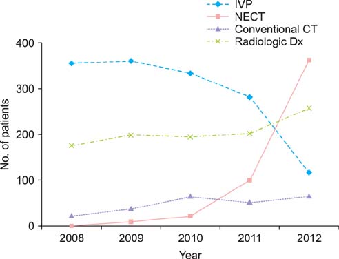
FIG. 1 Trend change in imaging use for patients with acute flank pain. Diamond curve indicates intravenous pyelogram (IVP). Squares indicate non-contrast-enhanced computed tomography (NECT). Triangles indicate conventional computed tomography (CT). Multiplication sign indicates radiologic diagnosis (Dx).
Reference
-
1. Smith RC, Rosenfield AT, Choe KA, Essenmacher KR, Verga M, Glickman MG, et al. Acute flank pain: comparison of non-contrast-enhanced CT and intravenous urography. Radiology. 1995; 194:789–794.2. Yilmaz S, Sindel T, Arslan G, Ozkaynak C, Karaali K, Kabaalioglu A, et al. Renal colic: comparison of spiral CT, US and IVU in the detection of ureteral calculi. Eur Radiol. 1998; 8:212–217.3. Niall O, Russell J, MacGregor R, Duncan H, Mullins J. A comparison of noncontrast computerized tomography with excretory urography in the assessment of acute flank pain. J Urol. 1999; 161:534–537.4. Spencer BA, Wood BJ, Dretler SP. Helical CT and ureteral colic. Urol Clin North Am. 2000; 27:231–241.5. Hyams ES, Korley FK, Pham JC, Matlaga BR. Trends in imaging use during the emergency department evaluation of flank pain. J Urol. 2011; 186:2270–2274.6. Larson DB, Johnson LW, Schnell BM, Salisbury SR, Forman HP. National trends in CT use in the emergency department: 1995-2007. Radiology. 2011; 258:164–173.7. Fielding JR, Steele G, Fox LA, Heller H, Loughlin KR. Spiral computerized tomography in the evaluation of acute flank pain: a replacement for excretory urography. J Urol. 1997; 157:2071–2073.8. Smith RC, Verga M, McCarthy S, Rosenfield AT. Diagnosis of acute flank pain: value of unenhanced helical CT. AJR Am J Roentgenol. 1996; 166:97–101.9. Ahmad NA, Ather MH, Rees J. Unenhanced helical computed tomography in the evaluation of acute flank pain. Int J Urol. 2003; 10:287–292.10. Song HJ, Cho ST, Kim KK. Investigation of the location of the ureteral stone and diameter of the ureter in patients with renal colic. Korean J Urol. 2010; 51:198–201.11. Katzberg RW. Urography into the 21st century: new contrast media, renal handling, imaging characteristics, and nephrotoxicity. Radiology. 1997; 204:297–312.12. Remer EM, Herts BR, Streem SB, Hesselink DP, Shiesly DA, Yost AJ, et al. Spiral noncontrast CT versus combined plain radiography and renal US after extracorporeal shock wave lithotripsy: cost-identification analysis. Radiology. 1997; 204:33–37.13. Brenner DJ, Hall EJ. Computed tomography: an increasing source of radiation exposure. N Engl J Med. 2007; 357:2277–2284.14. Fazel R, Krumholz HM, Wang Y, Ross JS, Chen J, Ting HH, et al. Exposure to low-dose ionizing radiation from medical imaging procedures. N Engl J Med. 2009; 361:849–857.15. Katz SI, Saluja S, Brink JA, Forman HP. Radiation dose associated with unenhanced CT for suspected renal colic: impact of repetitive studies. AJR Am J Roentgenol. 2006; 186:1120–1124.16. Sodickson A, Baeyens PF, Andriole KP, Prevedello LM, Nawfel RD, Hanson R, et al. Recurrent CT, cumulative radiation exposure, and associated radiation-induced cancer risks from CT of adults. Radiology. 2009; 251:175–184.17. Ferrandino MN, Bagrodia A, Pierre SA, Scales CD Jr, Rampersaud E, Pearle MS, et al. Radiation exposure in the acute and short-term management of urolithiasis at 2 academic centers. J Urol. 2009; 181:668–672.
- Full Text Links
-
- Actions
-
Cited
- CITED
-
- Close
- Share
-
- Similar articles
-
- Hounsfield Units of Urinary Calculi as a Predictor of the Therapeutic Effect of Extracorporeal Shockwave Lithotripsy
- Comparison of CT Urography and Intravenous Urography in Patients with Hematuria
- Massive Intraventricular Air Embolism after Contrast-enhanced CT: Report of Two Cases
- Decision Factors in Performance of Intravenous Contrast-enhanced Computed Tomography for Patients with Acute Flank Pain in an Emergency Department
- Transient Orbitofacial Angioedema due to Intravenous Iodinated Contrast Media During Computed Tomography: CT Findings
