Reversible Pulmonary Hypertension in Adolescent with Left Atrial Myxoma
- Affiliations
-
- 1Division of Cardiology, Daegu Catholic University Medical Center, Daegu, Korea. kks7379@cu.ac.kr
- 2Department of Internal Medicine, Daegu Catholic University Medical Center, Daegu, Korea.
- 3Department of Pediatrics, Daegu Catholic University Medical Center, Daegu, Korea.
- KMID: 1980388
- DOI: http://doi.org/10.4250/jcu.2011.19.4.221
Abstract
- We report a patient of left atrial huge myxoma presenting with severe pulmonary hypertension in adolescents. A patient was a 14-year-old boy presented with sudden onset dyspnea. Transthoracic echocardiographic study revealed the presence of a nodular, 4.34 x 8.11 cm sized, mobile, hyperechoic mass in the left atrium and severe pulmonary hypertension with tricuspid insufficiency. After surgical therapy, tricuspid regurgitation and pulmonary hypertension was decreased and the patient was stabilized and had an uneventful clinical course.
MeSH Terms
Figure
-

Fig. 1 Chest X-ray showed mild cardiomegaly and increased pulmonary vascular marking in both lungs.
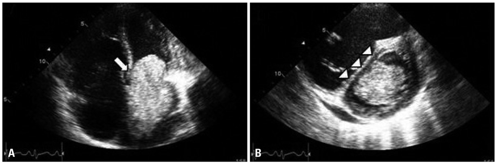
Fig. 2 A: Transthoracic echocardiography showed a 6 × 5 × 4.5 cm sized large left atrial mass (arrow) and right ventricular enlargement in apical 4 chamber view. B: D-shaped left ventricle during diastolic phase in parasternal short axis view (arrowhead).
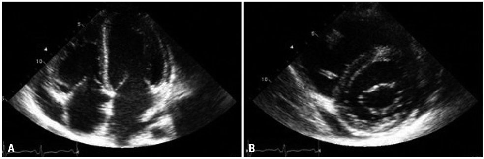
Fig. 3 A: Transthoracic echocardiography after mass removal showed a no visible left atrial mass in apical 4 chamber. B: No D-shaped left ventricle during diastolic phase in parasternal short axis view.

Fig. 4 Transthoracic doppler echocardiography showed tricuspid regurgitation with maximal pressure gradient (81.61 mm Hg).

Fig. 5 Transthoracic doppler echocardiography after mass removal showed decreased tricuspid regurgitation with maximal pressure gradient (39.37 mm Hg).

Fig. 6 Gross specimen of left atrial mass, friable hemorrhagic nodular mass, measuring 6 × 5 × 4.5 cm in size
Cited by 1 articles
-
Left Atrial Myxoma Mimicking Polyarteritis Nodosa
Gokhan Sargin, Taskin Senturk
Yonsei Med J. 2015;56(4):1165-1166. doi: 10.3349/ymj.2015.56.4.1165.
Reference
-
1. Kuon E, Kreplin M, Weiss W, Dahm JB. The challenge presented by right atrial myxoma. Herz. 2004. 29:702–709.
Article2. Reynen K. Cardiac myxomas. N Engl J Med. 1995. 333:1610–1617.
Article3. Pucci A, Gagliardotto P, Zanini C, Pansini S, di Summa M, Mollo F. Histopathologic and clinical characterization of cardiac myxoma: review of 53 cases from a single institution. Am Heart J. 2000. 140:134–138.
Article4. Vasquez A, Sethi G, Maximov M, Marcus FI. Atrial myxomas in the elderly: a case report and review of the literature. Am J Geriatr Cardiol. 2004. 13:39–44.
Article5. Goswami KC, Shrivastava S, Bahl VK, Saxena A, Manchanda SC, Wasir HS. Cardiac myxomas: clinical and echocardiographic profile. Int J Cardiol. 1998. 63:251–259.
Article6. Nakano T, Mayumi H, Hisahara M, Yasui H, Tokunaga K. The relationship between functional class, pulmonary artery pressure and size in left atrial myxoma. Cardiovasc Surg. 1996. 4:320–323.
Article
- Full Text Links
-
- Actions
-
Cited
- CITED
-
- Close
- Share
-
- Similar articles
-
- Successful Surgical Treatment of a Right Atrial Myxoma Complicated by Pulmonary Embolism
- Recurred Right Atrial Myxoma after Resection of Left Atrial Myxoma (Recurred Myxoma): A case report
- A Case of Left Atrial Myxoma Presenting as Acute Pulmonary Edema
- Silent Left Large Atrial Myxoma: A Patient with Serial Electrocardiogram Variation
- A Case of Multiple Right Atrial Myxomas with Pulmonary Embolism
