Right Coronary Cusp Prolapse Resembling Subpulmonic Stenosis in an Old Adult Patient with Ventricular Septal Defect
- Affiliations
-
- 1Cardiology Division, Heart Center, Gachon University Gil Hospital, Incheon, Korea. wjcheart@gmail.com
- 2Department of Cardiovascular Surgery, Gachon University Gil Hospital, Incheon, Korea.
- 3Gachon Cardiovascular Research Institute, Gachon University School of Medicine, Incheon, Korea.
- KMID: 1980387
- DOI: http://doi.org/10.4250/jcu.2011.19.4.216
Abstract
- Ventricular septal defect (VSD) can be associated with various complications such as aortic regurgitation (AR). AR in VSD come from a deficiency or hypoplasia of the conal septum which leads to abnormal apposition in diastole and prolapse of the poorly supported noncoronary or right coronary cusp through the VSD into the right ventricle resembling subpulmonic stenosis and subsequently results in distortion of the aortic valve and progressive AR. AR often increases in severity with age and it indicates a worse prognosis. Therefore, appropriate timing of surgical repair in progressive AR in VSD might be important. Until now, many earlier experiences about surgical repair of AR complicating VSD were on adolescents or young adults. We reported a case of AR in 48-year-old male patient with right coronary cusp prolapse complicating the subarterial type of VSD which was properly assessed by echocardiography and was successfully treated with surgical repair. Right coronary cusp or noncoronary cusp prolapse should be suspected in AR complicating VSD through proper echocardiographic assessment and the surgical repair on VSD and distorted aortic valve should be considered in the old patient, as well as the young.
MeSH Terms
Figure
-
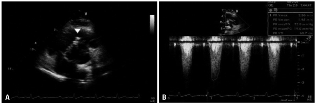
Fig. 1 Transthoracic echocardiography with the short-axis view at the aortic level (A) with continuous wave Doppler at the right ventricular outflow tract (RVOT) (B). It showed mild RVOT obstruction resembling subpulmonic stenosis (arrowhead) with increased peak and mean pressure gradients (32 mm Hg and 19 mm Hg, respectively).
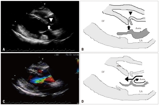
Fig. 2 The transthoracic parasternal long axis views on admission with illustrations. A and B: There were a discontinuity on interventricular septum showing subarterial type ventricular septal defect (VSD, arrow) and a finger-like projection of sinus of Valsalva (arrowhead) resulting from longstanding high velocity jet through VSD. C: Severe aortic regurgitant jet was identified with color Doppler. D: A section of the aortic root illustration showed the direction of blood flow (arrows). LV: left ventricle, LA: left atrium, IVS: interventricular septum.
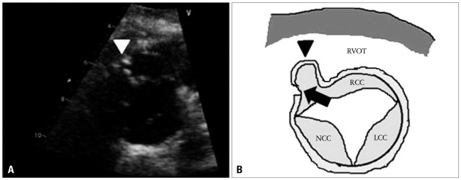
Fig. 3 The transthoracic short-axis view at the aortic level on admission (A) and illustration (B). The right coronary cusp and sinus of Valsalva are driven into the right ventricle (RV) due to longstanding high velocity jet produced by the left-to-right shunt (arrow). As the result, the right ventricular outflow tract obstruction occurred (arrowhead). RCC: right coronary cusp, NCC: noncoronary cusp, LCC: left coronary cusp, RVOT: right ventricular outflow tract.
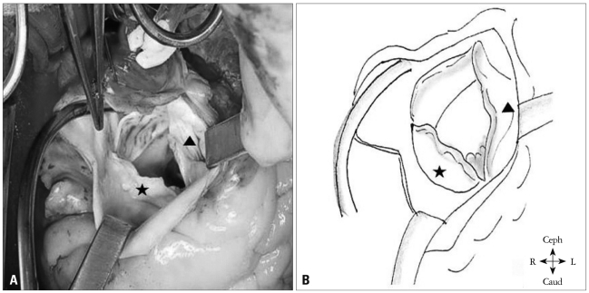
Fig. 4 Surgical findings of the aortic valve via aortotomy (A) and illustration (B). It showed a retracted right coronary cusp (RCC) (arrowhead) and the commissural fusion and calcification between the RCC and the noncoronary cusp (star).
Reference
-
1. Ammash NM, Warnes CA. Ventricular septal defects in adults. Ann Intern Med. 2001; 135:812–824. PMID: 11694106.2. Soto B, Becker AE, Moulaert AJ, Lie JT, Anderson RH. Classification of ventricular septal defects. Br Heart J. 1980; 43:332–343. PMID: 7437181.3. Ueda M, Fujimoto T, Becker AE. The infundibular septum in normal hearts and in hearts with isolated ventricular septal defect. A comparison between Japanese and Dutch hearts. Jpn Heart J. 1986; 27:635–643. PMID: 3820574.4. Elgamal MA, Hakimi M, Lyons JM, Walters HL 3rd. Risk factors for failure of aortic valvuloplasty in aortic insufficiency with ventricular septal defect. Ann Thorac Surg. 1999; 68:1350–1355. PMID: 10543505.5. Kidd L, Driscoll DJ, Gersony WM, Hayes CJ, Keane JF, O'Fallon WM, Pieroni DR, Wolfe RR, Weidman WH. Second natural history study of congenital heart defects. Results of treatment of patients with ventricular septal defects. Circulation. 1993; 87:I38–I51. PMID: 8425321.6. Neumayer U, Stone S, Somerville J. Small ventricular septal defects in adults. Eur Heart J. 1998; 19:1573–1582. PMID: 9820997.7. Gabriel HM, Heger M, Innerhofer P, Zehetgruber M, Mundigler G, Wimmer M, Maurer G, Baumgartner H. Long-term outcome of patients with ventricular septal defect considered not to require surgical closure during childhood. J Am Coll Cardiol. 2002; 39:1066–1071. PMID: 11897452.8. Lue HC, Sung TC, Hou SH, Wu MH, Cheng SJ, Chu SH, Hung CR. Ventricular septal defect in Chinese with aortic valve prolapse and aortic regurgitation. Heart Vessels. 1986; 2:111–116. PMID: 3759798.9. Mori K, Matsuoka S, Tatara K, Hayabuchi Y, Nii M, Kuroda Y. Echocardiographic evaluation of the development of aortic valve prolapse in supracristal ventricular septal defect. Eur J Pediatr. 1995; 154:176–181. PMID: 7758512.10. Sohn DW. Symptom based echocardiographic approach: dyspnea. J Korean Soc Echocardiogr. 2004; 12:5–9.11. Pieroni DR, Nishimura RA, Bierman FZ, Colan SD, Kaufman S, Sanders SP, Seward JB, Tajik AJ, Wiggins JW, Zahka KG. Second natural history study of congenital heart defects. Ventricular septal defect: echocardiography. Circulation. 1993; 87:I80–I88. PMID: 8425326.12. Garg A, Shrivastava S, Radhakrishnan S, Dev V, Saxena A. Doppler assessment of interventricular pressure gradient across isolated ventricular septal defect. Clin Cardiol. 1990; 13:717–721. PMID: 2257713.13. Schmidt KG, Cassidy SC, Silverman NH, Stanger P. Doubly committed subarterial ventricular septal defects: echocardiographic features and surgical implications. J Am Coll Cardiol. 1988; 12:1538–1546. PMID: 3057035.14. Backer CL, Idriss FS, Zales VR, Ilbawi MN, DeLeon SY, Muster AJ, Mavroudis C. Surgical management of the conal (supracristal) ventricular septal defect. J Thorac Cardiovasc Surg. 1991; 102:288–295. discussion 295-6. PMID: 1865702.15. Momma K, Toyama K, Takao A, Ando M, Nakazawa M, Hirosawa K, Imai Y. Natural history of subarterial infundibular ventricular septal defect. Am Heart J. 1984; 108:1312–1317. PMID: 6496290.16. Mahoney LT. Acyanotic congenital heart disease. Atrial and ventricular septal defects, atrioventricular canal, patent ductus arteriosus, pulmonic stenosis. Cardiol Clin. 1993; 11:603–616. PMID: 8252562.17. Menahem S, Johns JA, del Torso S, Goh TH, Venables AW. Evaluation of aortic valve prolapse in ventricular septal defect. Br Heart J. 1986; 56:242–249. PMID: 3756041.18. Leung MP, Beerman LB, Siewers RD, Bahnson HT, Zuberbuhler JR. Long-term follow-up after aortic valvuloplasty and defect closure in ventricular septal defect with aortic regurgitation. Am J Cardiol. 1987; 60:890–894. PMID: 3661405.19. Otterstad JE, Erikssen J, Frøysaker T, Simonsen S. Long term results after operative treatment of isolated ventricular septal defect in adolescents and adults. Acta Med Scand Suppl. 1986; 708:1–39. PMID: 3461690.20. Ellis JH 4th, Moodie DS, Sterba R, Gill CC. Ventricular septal defect in the adult: natural and unnatural history. Am Heart J. 1987; 114:115–120. PMID: 3604855.21. Gersony WM, Hayes CJ, Driscoll DJ, Keane JF, Kidd L, O'Fallon WM, Pieroni DR, Wolfe RR, Weidman WH. Second natural history study of congenital heart defects. Quality of life of patients with aortic stenosis, pulmonary stenosis, or ventricular septal defect. Circulation. 1993; 87:I52–I65. PMID: 8425323.22. Warnes CA, Williams RG, Bashore TM, Child JS, Connolly HM, Dearani JA, del Nido P, Fasules JW, Graham TP Jr, Hijazi ZM, Hunt SA, King ME, Landzberg MJ, Miner PD, Radford MJ, Walsh EP, Webb GD, Smith SC Jr, Jacobs AK, Adams CD, Anderson JL, Antman EM, Buller CE, Creager MA, Ettinger SM, Halperin JL, Hunt SA, Krumholz HM, Kushner FG, Lytle BW, Nishimura RA, Page RL, Riegel B, Tarkington LG, Yancy CW. American College of Cardiology. American Heart Association Task Force on Practice Guidelines (Writing Committee to Develop Guidelines on the Management of Adults With Congenital Heart Disease). American Society of Echocardiography. Heart Rhythm Society. International Society for Adult Congenital Heart Disease. Society for Cardiovascular Angiography and Interventions. Society of Thoracic Surgeons. ACC/AHA 2008 guidelines for the management of adults with congenital heart disease: a report of the American College of Cardiology/American Heart Association Task Force on Practice Guidelines (Writing Committee to Develop Guidelines on the Management of Adults With Congenital Heart Disease). Developed in Collaboration With the American Society of Echocardiography, Heart Rhythm Society, International Society for Adult Congenital Heart Disease, Society for Cardiovascular Angiography and Interventions, and Society of Thoracic Surgeons. J Am Coll Cardiol. 2008; 52:e1–e121. PMID: 19038677.23. Mongeon FP, Burkhart HM, Ammash NM, Dearani JA, Li Z, Warnes CA, Connolly HM. Indications and outcomes of surgical closure of ventricular septal defect in adults. JACC Cardiovasc Interv. 2010; 3:290–297. PMID: 20298987.
- Full Text Links
-
- Actions
-
Cited
- CITED
-
- Close
- Share
-
- Similar articles
-
- Hemodynamic Study of Subpulmonic Ventricular Defect(by Cardiac Catheterization and Cineangiocardiography)
- A Case of Ruptured Aneurysm of Right Sinus of Valsalva Diagnosed Preoperatively by Echocardiography
- Two Cases of Ventricular Septal Defect with Aortic Insufficiency
- Subpulmonic Ventricular Septal Defect with Aortic Insufficiency
- A Case Report of Double Outlet Right Ventricle(S.D.L.) with Subpulmonic Ventricular Septal Defect and Pulmonary Stenosis
