Prominent Crista Terminalis in Patients with Embolic Events
- Affiliations
-
- 1Cardiovascular Center, Korea University Guro Hospital, Korea University College of Medicine, Seoul, Korea. withnoel@hanmail.net
- 2Cardiothoracic Division, Department of Radiology, Korea University Guro Hospital, Korea University College of Medicine, Seoul, Korea.
- KMID: 1980372
- DOI: http://doi.org/10.4250/jcu.2011.19.3.156
Abstract
- A prominent crista terminalis is a normal anatomic variant which consist of thick muscular bridge within the right atrium. However, it could be often misdiagnosed with an abnormal mass on the transthoracic echocardiography. The case report presented here, describe the findings of transthoracic echocardiography that suggested a right atrial mass in patients with pulmonary embolism. However, subsequent transesophageal echocardiography and cardiac computed tomography/magnetic resonance imaging differentiated a true right atrial mass from a prominent crista terminalis.
Keyword
Figure
-
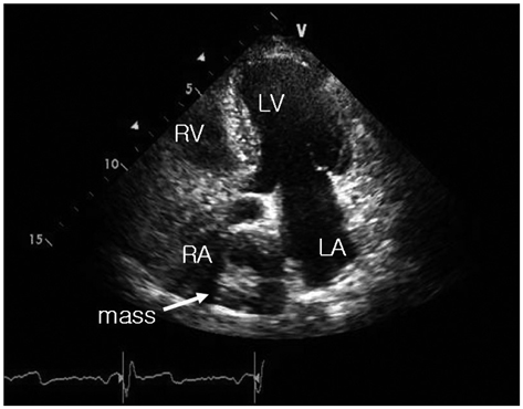
Fig. 1 Transthoracic echocardiogram reveals an echogenic non-mobile mass in the right atrium. LA: left atrium, LV: left ventricle, RA: right atrium, RV: right ventricle.
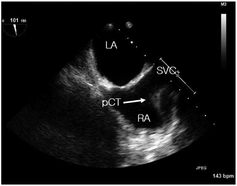
Fig. 2 Transesophageal echocardiogram shows a prominent crista terminalis at the superior part of right atrium close to superior vena cava. LA: left atrium, pCT: prominent crista terminalis, RA: right atrium, SVC: superior vena cava.
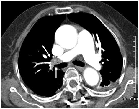
Fig. 3 A very large pulmonary embolism is seen in the right pulmonary artery.
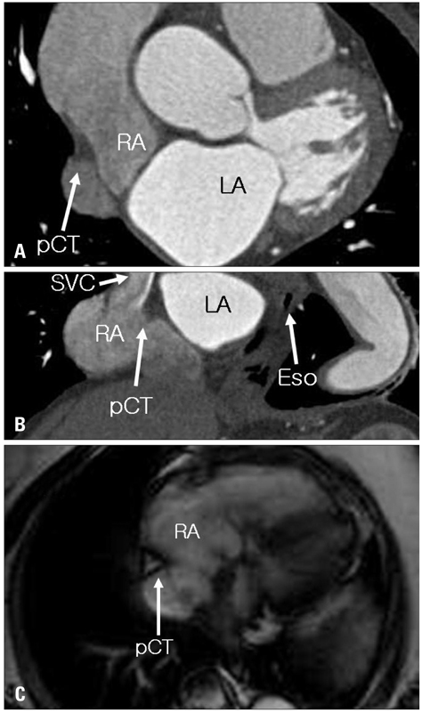
Fig. 4 Chest computed tomography (CT) (A and B) and magnetic resonance imaging (C) reveal a prominent crista terminalis without any definite mass in the right atrium. Eso: esophagus, LA: left atrium, pCT: prominent crista terminalis, RA: right atrium, SVC: superior vena cava.
Reference
-
1. Meier RA, Hartnell GG. MRI of right atrial pseudomass: is it really a diagnostic problem? J Comput Assist Tomogr. 1994. 18:398–401.2. Pharr JR, West MB, Kusumoto FM, Figueredo VM. Prominent crista terminalis appearing as a right atrial mass on transthoracic echocardiogram. J Am Soc Echocardiogr. 2002. 15:753–755.
Article3. Pharr JR, Figueredo VM. Lipomatus hypertrophy of the atrial septum and prominent crista terminalis appearing as a right atrial mass. Eur J Echocardiogr. 2002. 3:159–161.
Article4. McKay T, Thomas L. Prominent crista terminalis and Eustachian ridge in the right atrium: two dimensional (2D) and three dimensional (3D) imaging. Eur J Echocardiogr. 2007. 8:288–291.
Article5. Gaudio C, Di Michele S, Cera M, Nguyen BL, Pannarale G, Alessandri N. Prominent crista terminalis mimicking a right atrial mixoma: cardiac magnetic resonance aspects. Eur Rev Med Pharmacol Sci. 2004. 8:165–168.6. D'Amato N, Pierfelice O, D'Agostino C. Crista terminalis bridge: a rare variant mimicking right atrial mass. Eur J Echocardiogr. 2009. 10:444–445.
- Full Text Links
-
- Actions
-
Cited
- CITED
-
- Close
- Share
-
- Similar articles
-
- Nervus terminalis and nerves to the vomeronasal organ: a study using human fetal specimens
- The Clinical Impacts of Apparent Embolic Event and the Predictors of In-Hospital Mortality in Patients with Infective Endocarditis
- Acromegaloid Facial Appearance Syndrome with Generalized Hypertrichosis Terminalis
- Developmental Changes of Glial Fibrillary Acidic Protein (GFAP) and Proliferating Cell Nuclear Antigen (PCNA) Immunoreactivity of the Ependyma lining the Central Canal and Ventriculus Terminalis in the Human Fetus
- Comparison of Fornix and Stria Terminalis Connectivity among First-Episode Schizophrenia, Chronic Schizophrenia and Healthy Controls
