A Case of Fungating Type Natural Killer Like T Cell Lymphoma of the Ascending Colon
- Affiliations
-
- 1Department of Internal Medicine and Research Institute for Convergence of Biomedical Science and Technology, Pusan National University Yangsan Hospital, Pusan National University School of Medicine, Yangsan, Korea. psubumi@naver.com
- 2Department of Pathology, Pusan National University Yangsan Hospital, Pusan National University School of Medicine, Yangsan, Korea.
- KMID: 1979208
- DOI: http://doi.org/10.4166/kjg.2014.64.4.229
Abstract
- Primary colorectal lymphoma is a very rare disease entity that accounts for less than 0.2-0.65% of all colon cancers. It is as an extranodal lymphoma of the colon that mainly arises from B cells and primary colorectal lymphoma that arises from T cells is very rare both in Western countries and in Korea. Colonic lymphoma can be classified endoscopically into 5 categories as follows: fungating, ulcerative, infiltrative, ulcerofungating, and ulceroinfiltrative type. The endoscopic features of primary colorectal lymphoma differ according to their cellular origin; about half of B cell lymphomas are fungating type whereas most of T cell lymphomas are of ulcerative or ulceroinfiltrative type. Mass forming primary T cell lymphoma of the colon is extremely rare. Herein, we present a case of primary natural killer like T cell lymphoma of the colon presenting as fungating type with review of literature.
MeSH Terms
Figure
-

Fig. 1. Abdominal CT scan findings. (A) CT scan performed 3 months ago shows no evidence of abnormality on the hepatic flexure. (B) CT scan taken at admission shows newly developed huge mass (arrow) with pericolic fat stranding on the hepatic flexure.

Fig. 2. Endoscopic finding. A large fungating mass is seen on distal ascending colon which almost obstructs the bowel lumen.

Fig. 3. Microscopic findings of the biopsy specimens from the colon mass. (A) Diffuse infiltration of medium-sized atypical lymphoid cells with irregular, pleomorphic, hyperchromatic or vesicular nuclei and many mitotic figures are observed (H&E, ×400). (B) Immunohistochemical stain shows that lymphoma cells are CD3 positive T lymphocytes (×200) that are positive for (C) CD 56 (×200) and (D) CD 8 (×200).
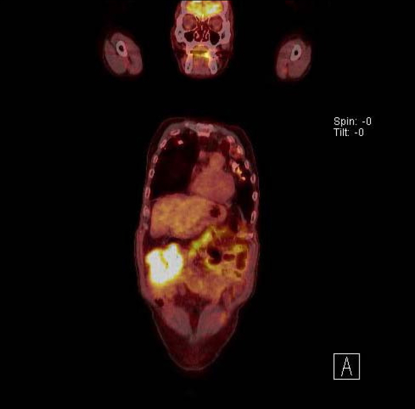
Fig. 4. PET scan finding. The mass at hepatic flexure shows intense hypermetabolism with standardized uptake value of 10.8.
Reference
-
References
1. Chan JK. Gastrointestinal lymphomas: an overview with emphasis on new findings and diagnostic problems. Semin Diagn Pathol. 1996; 13:260–296.2. Shepherd NA, Hall PA, Coates PJ, Levison DA. Primary malignant lymphoma of the colon and rectum. A histopathological and immunohistochemical analysis of 45 cases with clinicopathological correlations. Histopathology. 1988; 12:235–252.
Article3. Kim YH, Lee JH, Yang SK, et al. Primary colon lymphoma in Korea: a KASID (Korean Association for the Study of Intestinal Diseases) Study. Dig Dis Sci. 2005; 50:2243–2247.
Article4. Kim HS, Lee DK, Baik SK, Kwon SO, Cho MY, Ko YH. Primary CD56+ T/NK cell lymphoma of the colon. J Gastroenterol. 2002; 37:939–946.
Article5. Dawson IM, Cornes JS, Morson BC. Primary malignant lymphoid tumours of the intestinal tract. Report of 37 cases with a study of factors influencing prognosis. Br J Surg. 1961; 49:80–89.
Article6. Jaffe ES. The 2008 WHO classification of lymphomas: implications for clinical practice and translational research. Hematology Am Soc Hematol Educ Program;2009. p. 523–531.7. Harris NL, Jaffe ES, Diebold J, et al. The World Health Organization classification of neoplasms of the hematopoietic and lymphoid tissues: report of the Clinical Advisory Committee meeting–Airlie House, Virginia, November, 1997. Hematol J. 2000; 1:53–66.8. Sarris A, Ford R. Recent advances in the molecular pathogenesis of lymphomas. Curr Opin Oncol. 1999; 11:351–363.
Article9. Kwong YL. Natural killer-cell malignancies: diagnosis and treatment. Leukemia. 2005; 19:2186–2194.
Article10. Jaffe ES. Classification of natural killer (NK) cell and NK-like T-cell malignancies. Blood. 1996; 87:1207–1210.11. Tung CL, Hsieh PP, Chang JH, Chen RS, Chen YJ, Wang JS. Intestinal T-cell and natural killer-cell lymphomas in Taiwan with special emphasis on 2 distinct cellular types: natural killer-like cytotoxic T cell and true natural killer cell. Hum Pathol. 2008; 39:1018–1025.
Article12. Tse E, Gill H, Loong F, et al. Type II enteropathy-associated T-cell lymphoma: a multicenter analysis from the Asia Lymphoma Study Group. Am J Hematol. 2012; 87:663–668.
Article13. Ko YH, Cho EY, Kim JE, et al. NK and NK-like T-cell lymphoma in extranasal sites: a comparative clinicopathological study according to site and EBV status. Histopathology. 2004; 44:480–489.
Article14. Chim CS, Au WY, Shek TW, et al. Primary CD56 positive lymphomas of the gastrointestinal tract. Cancer. 2001; 91:525–533.
Article15. Zighelboim J, Larson MV. Primary colonic lymphoma. Clinical presentation, histopathologic features, and outcome with com-bination chemotherapy. J Clin Gastroenterol. 1994; 18:291–297.16. Lee YJ, Lee JH. Gastrointestinal lymphoma. Korean J Helicobacter Up Gastrointest Res. 2012; 12:158–165.
Article17. Zheng S, Ouyang Q, Li G, et al. Primary intestinal NK/T cell lymphoma: a clinicopathologic study of 25 Chinese cases. Arch Iran Med. 2012; 15:36–42.18. Chun HB, Baek IH, Lee MS, et al. Jejunocolic fistula associated with an intestinal T cell lymphoma. Gut Liver. 2011; 5:387–390.
Article19. Ryu SH, Cheon JH, Kim JY, et al. The early diagnostic accuracy for gastrointestinal T-cell lymphoma from a perspective of gas-troenterologist. Intest Res. 2011; 9:19–26.20. Lister TA, Crowther D, Sutcliffe SB, et al. Report of a committee convened to discuss the evaluation and staging of patients with Hodgkin's disease: Cotswolds meeting. J Clin Oncol. 1989; 7:1630–1636.
Article
