Evaluation of hyoid bone movements in subjects with open bite: a study with real-time balanced turbo field echo cine-magnetic resonance imaging
- Affiliations
-
- 1Section of Orthodontics, Gulhane Military Medical Academy, Haydarpasha Education Hospital, Istanbul, Turkey. kseniz@yahoo.com
- 2Department of Orthodontics, Gulhane Military Medical Academy, Ankara, Turkey.
- KMID: 1976684
- DOI: http://doi.org/10.4041/kjod.2012.42.6.318
Abstract
OBJECTIVE
To assess the position and movements of the hyoid bone during deglutition in patients with open bite.
METHODS
Thirty-six subjects were divided into 2 groups according to the presence of anterior open bite. The open bite group (OBG) and control group each comprised 18 patients with a mean overbite of -4.9 +/- 1.9 mm and 1.9 +/- 0.7 mm. The position of the hyoid bone during the 4 stages of deglutition was evaluated by measuring vertical and horizontal movement of the bone.
RESULTS
Interactions of group and stage showed no significant effect on the measurements (p > 0.05). However, when group and stage were evaluated individually, they showed significant effects on the measurements (p < 0.001). In OBG, the hyoid bone was more inferiorly and posteriorly positioned, and this position continued during the deglutition stages.
CONCLUSIONS
The hyoid bone reaches the maximum anterior position at the oral stage and maximum superior position at the pharyngeal stage during deglutition. Open bite does not change the displacement pattern of the bone during deglutition. The hyoid bone is positioned more inferiorly and posteriorly in patients with open bite because of released tension on the suprahyoid muscles.
Figure
-
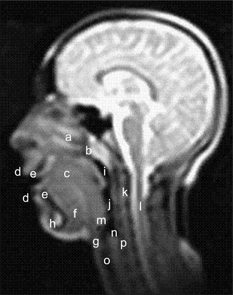
Figure 1 Anatomical structures visible on cine magnetic resonance imaging (a) nasal cavity, (b) nasopharynx, (c) tongue, (d) lips, (e) incisors, (f) floor of the mouth, (g) hyoid bone, (h) symphysis, (i) pharynx, (j) larynx, (k) spinal column, (l) spinal cord, (m) epiglottis, (n) cricoid cartilage and cricotracheal membrane, (o) trachea, and (p) upper portion of the esophagus.
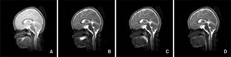
Figure 2 Stages of deglutition. A, stage 1 (oral preparatory stage), the tongue tip contacts the maxillary incisors and/or the palatal mucosa; B, stage 2 (oral stage), the dorsum of the tongue loses contact with the soft palate; C, stage 3 (pharyngeal stage), the bolus crosses the posterior or inferior margin of the mandibular ramus; and D, stage 4 (esophageal stage), the bolus enters the esophageal opening.
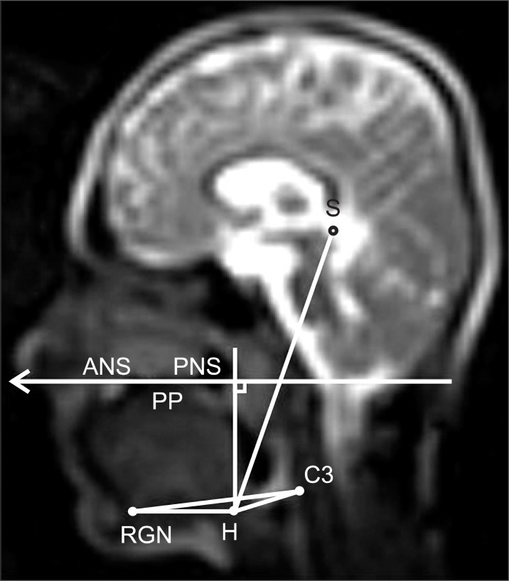
Figure 3 Measurements used for evaluating hyoid bone movements. (1) H-S, distance between hyoidale (H, the most anterosuperior point of the hyoid bone) and point sella (S); (2) H-PP, perpendicular distance of hyoidale to the palatal plane (PP); (3) H-C3RGN, perpendicular distance of hyoidale to the line connecting the most anteroinferior point of the third cervical vertebra (C3) and retrognathion (RNG, the most prominent point of the posterior border of the symphysis); (4) H-C3, distance between hyoidale and the most anteroinferior point of the third cervical vertebra; and (5) H-RGN, distance between hyoidale and retrognathion.
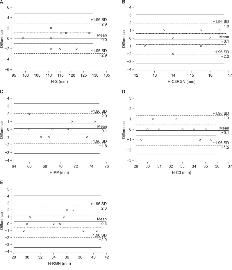
Figure 4 Bland-Altman plots of the average differences in the measurements. A, H-S; B, H-C3RGN; C, H-PP; D, H-C3; and E, H-RGN. The overall bias of each measurement is shown, along with the 95% agreement interval. SD, Standard deviation. Refer to Figure 3 for measurements and abbreviations.
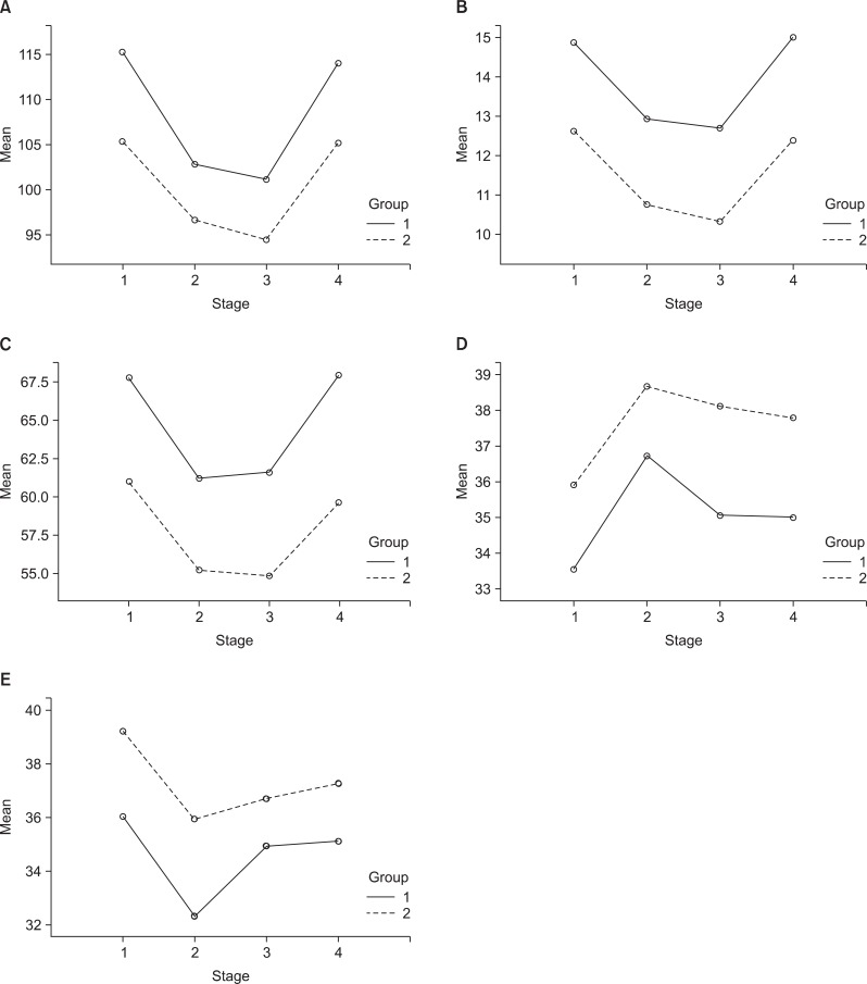
Figure 5 Plots of the mean A, H-S; B, H-C3RGN; C, H-PP; D, H-C3; and E, H-RGN values as a function of the stages of deglutition in the open bite group (group 1) and the control group (group 2). Refer to Figure 3 for measurements and abbreviations.
Reference
-
1. Rocabado M. Biomechanical relationship of the cranial, cervical, and hyoid regions. J Craniomandibular Pract. 1983; 1:61–66. PMID: 6586872.
Article2. Hayes RJ, Sarver DM, Jacobson A. The quantification of soft tissue cervicomental changes after mandibular advancement surgery. Am J Orthod Dentofacial Orthop. 1994; 105:383–391. PMID: 8154464.
Article3. Güven O, Saraçoğlu U. Changes in pharyngeal airway space and hyoid bone positions after body ostectomies and sagittal split ramus osteotomies. J Craniofac Surg. 2005; 16:23–30. PMID: 15699641.
Article4. Marşan G, Oztaş E, Cura N, Kuvat SV, Emekli U. Changes in head posture and hyoid bone position in Turkish Class III patients after mandibular setback surgery. J Craniomaxillofac Surg. 2010; 38:113–121. PMID: 19447640.
Article5. Gobeille DM, Bowman DC. Hyoid and muscle changes following distal repositioning of the tongue. Am J Orthod. 1976; 70:282–289. PMID: 1066966.
Article6. Adamidis IP, Spyropoulos MN. The effects of lymphadenoid hypertrophy on the position of the tongue, the mandible and the hyoid bone. Eur J Orthod. 1983; 5:287–294. PMID: 6581051.
Article7. Tallgren A, Solow B. Hyoid bone position, facial morphology and head posture in adults. Eur J Orthod. 1987; 9:1–8. PMID: 3470181.
Article8. Behlfelt K, Linder-Aronson S, Neander P. Posture of the head, the hyoid bone, and the tongue in children with and without enlarged tonsils. Eur J Orthod. 1990; 12:458–467. PMID: 2086266.
Article9. Haralabakis NB, Toutountzakis NM, Yiagtzis SC. The hyoid bone position in adult individuals with open bite and normal occlusion. Eur J Orthod. 1993; 15:265–271. PMID: 8405131.
Article10. Fromm B, Lundberg M. Postural behaviour of the hyoid bone in normal occlusion and before and after surgical correction of mandibular protrusion. Sven Tandlak Tidskr. 1970; 63:425–433. PMID: 5271184.12. Andersen WS. The relationship of the tongue-thrust syndrome to maturation and other factors. Am J Orthod. 1963; 49:264–275.
Article13. Subtelny JD, Sakuda M. Open bite: diagnosis and treatment. Am J Orthod. 1964; 50:337–358.14. Mays KA, Palmer JB, Kuhlemeier KV. Influence of craniofacial morphology on hyoid movement: a preliminary correlational study. Dysphagia. 2009; 24:71–76. PMID: 18716836.
Article15. Sloan RF, Bench RW, Mulick JF, Ricketts RM, Brummett SW, Westover JL. The application of cephalometrics to cinefluorography: comparative analysis of hyoid movement patterns during deglutition in Class I and Class II orthodontic patients. Angle Orthod. 1967; 37:26–34. PMID: 5225691.16. Fujiki T, Takano-Yamamoto T, Noguchi H, Yamashiro T, Guan G, Tanimoto K. A cineradiographic study of deglutitive tongue movement and nasopharyngeal closure in patients with anterior open bite. Angle Orthod. 2000; 70:284–289. PMID: 10961777.17. Fujiki T, Inoue M, Miyawaki S, Nagasaki T, Tanimoto K, Takano-Yamamoto T. Relationship between maxillofacial morphology and deglutitive tongue movement in patients with anterior open bite. Am J Orthod Dentofacial Orthop. 2004; 125:160–167. PMID: 14765053.
Article18. Akin E, Sayin MO, Karaçay S, Bulakbaşi N. Real-time balanced turbo field echo cine-magnetic resonance imaging evaluation of tongue movements during deglutition in subjects with anterior open bite. Am J Orthod Dentofacial Orthop. 2006; 129:24–28. PMID: 16443474.
Article19. Yılmaz F, Sağdıç D, Karaçay S, Akin E, Bulakbası N. Tongue movements in patients with skeletal Class II malocclusion evaluated with real-time balanced turbo field echo cine magnetic resonance imaging. Am J Orthod Dentofacial Orthop. 2011; 139:e415–e425. PMID: 21536183.
Article20. Cunningham JB, McCrum-Gardner E. Power, effect and sample size using GPower: practical issues for researchers and members of research ethics committees. Evid Based Midwifery. 2007; 5:132–136.21. Sivarao DV, Mehendale HM. 2-Butoxyethanol autoprotection is due to resiliance of newly formed erythrocytes to hemolysis. Arch Toxicol. 1995; 69:526–532. PMID: 8534195.
Article22. Palmer JB, Rudin NJ, Lara G, Crompton AW. Coordination of mastication and swallowing. Dysphagia. 1992; 7:187–200. PMID: 1308667.
Article23. Karaçay S, Akin E, Sayin MO, Bulakbaşi N. Real time balanced turbo field echo Cine-MRI in the analysis of deglutition events and transit times. J Oral Rehabil. 2006; 33:646–653. PMID: 16922737.
Article24. Görgülü S, Sağdıç D, Akin E, Karaçay S, Bulakbası N. Tongue movements in patients with skeletal Class III malocclusions evaluated with real-time balanced turbo field echo cine magnetic resonance imaging. Am J Orthod Dentofacial Orthop. 2011; 139:e405–e414. PMID: 21536182.
Article25. Karacay S, Akin E, Ortakoglu K, Bengi AO. Dynamic MRI evaluation of tongue posture and deglutitive movements in a surgically corrected open bite. Angle Orthod. 2006; 76:1057–1065. PMID: 17090175.
Article26. Anagnostara A, Stoeckli S, Weber OM, Kollias SS. Evaluation of the anatomical and functional properties of deglutition with various kinetic high-speed MRI sequences. J Magn Reson Imaging. 2001; 14:194–199. PMID: 11477680.
Article27. Dodds WJ, Man KM, Cook IJ, Kahrilas PJ, Stewart ET, Kern MK. Influence of bolus volume on swallow-induced hyoid movement in normal subjects. AJR Am J Roentgenol. 1988; 150:1307–1309. PMID: 3259369.
Article28. Grabber TM, Vanarsdall RL, Vig K. Orthodontics: Current principals and techniques. 2005. 4th ed. St. Louis: Mosby.
- Full Text Links
-
- Actions
-
Cited
- CITED
-
- Close
- Share
-
- Similar articles
-
- Relationship between Class III malocclusion and hyoid bone displacement during swallowing: a cine-magnetic resonance imaging study
- The Comparison between Single Shot Turbo Spin Echo and B-FFE (Balanced Turbo Field-echo) in the Differentiation of Focal Liver Lesions
- Utility of Two Types of MR Cisternography for Patency Evaluation of Aqueduct and Third Ventriculostomy Site: Three Dimensional Sagittal Fast Spin Echo Sequence and Steady-State Coherent Fast Gradient Echo Sequence
- A radiographic study of the hyoid bone position in malocclusion
- Retrospective Electrocardiography-Gated Real-Time Cardiac Cine MRI at 3T: Comparison with Conventional Segmented Cine MRI
