Does hyrax expansion therapy affect maxillary sinus volume? A cone-beam computed tomography report
- Affiliations
-
- 1Department of Orthodontics, School of Dentistry, University of Texas Health Science Center at Houston, Houston, USA. sercan.akyalcin@uth.tmc.edu
- 2Department of Orthodontics, School of Dentistry, University of Alabama at Birmingham, Birmingham, USA.
- KMID: 1974413
- DOI: http://doi.org/10.5624/isd.2012.42.2.83
Abstract
- PURPOSE
The aim of this study was to investigate the initial effects of maxillary expansion therapy with Hyrax appliance and to evaluate the related changes in maxillary sinus volume.
MATERIALS AND METHODS
Thirty patients (20 females, 10 males; 13.8 years) requiring maxillary expansion therapy, as part of their comprehensive orthodontic treatment, were examined. Each patient had cone-beam computed tomography (CBCT) images taken before (T1) and after (T2) maxillary expansion therapy with a banded Hyrax appliance. Multiplanar slices were used to measure linear dimensions and palatal vault angle. Volumetric analysis was used to measure maxillary sinus volumes. Student t tests were used to compare the pre- and post-treatment measurements. Additionally, differences between two age groups were compared with Mann-Whitney U test. The level of significance was set at p=0.05.
RESULTS
Comparison of pre-treatment to post-treatment variables revealed significant changes in the transverse dimension related to both maxillary skeletal and dental structures and palatal vault angle, resulting in a widened palatal vault (p<0.05). Hard palate showed no significant movement in the vertical and anteroposterior planes. Nasal cavity width increased on a mean value of 0.93mm(SD=0.23, p<0.05). Maxillary sinus volume remained virtually stable. No significant age differences were observed in the sample.
CONCLUSION
Hyrax expansion therapy did not have a significant impact on maxillary sinus volume.
MeSH Terms
Figure
-

Fig. 1 A. Dental width measurements are carried out using both premolars and the canine. B. Palatal vault angle measurement is made at a point coronally at the palatal root of the first molars.
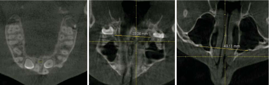
Fig. 2 The width of the incisive canal (A), the greater palatine foramen to contralateral greater palatine foramen distance (B), the infraorbital foramen to contralateral infraorbital foramen distance (C) are recorded on the axial images that are created perpendicular to the midsagittal plane for each of the anatomic landmark.
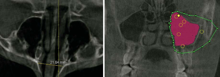
Fig. 3 A. Nasal cavity width is made on axial images. B. Boundary lines are drawn surrounding the sinus cavity in the axial, coronal, and sagittal views individually to help assist with the maxillary sinus volume measurement.
Reference
-
1. Haas AJ. Long-term posttreatment evaluation of rapid palatal expansion. Angle Orthod. 1980. 50:189–217.2. Bishara SE, Staley RN. Maxillary expansion: clinical implications. Am J Orthod Dentofacial Orthop. 1987. 91:3–14.
Article3. Adkins MD, Nanda RS, Currier GF. Arch perimeter changes on rapid palatal expansion. Am J Orthod Dentofacial Orthop. 1990. 97:194–199.
Article4. Geran RG, McNamara JA Jr, Baccetti T, Franchi L, Shapiro LM. A prospective long-term study on the effects of rapid maxillary expansion in the early mixed dentition. Am J Orthod Dentofacial Orthop. 2006. 129:631–640.
Article5. Baccetti T, Franchi L, Cameron CG, McNamara JA Jr. Treatment timing for rapid maxillary expansion. Angle Orthod. 2001. 71:343–350.6. Oliveira De Felippe NL, Da Silveira AC, Viana G, Kusnoto B, Smith B, Evans CA. Relationship between rapid maxillary expansion and nasal cavity size and airway resistance: short- and long-term effects. Am J Orthod Dentofacial Orthop. 2008. 134:370–382.
Article7. Timms DJ, Preston CB, Daly PF. A computed tomographic assessment of maxillary movement induced by rapid expansion - a pilot study. Eur J Orthod. 1982. 4:123–127.
Article8. Kau CH, Richmond S, Palomo JM, Hans MG. Three-dimensional cone beam computerized tomography in orthodontics. J Orthod. 2005. 32:282–293.9. Garrett BJ, Caruso JM, Rungcharassaeng K, Farrage JR, Kim JS, Taylor GD. Skeletal effects to the maxilla after rapid maxillary expansion assessed with cone-beam computed tomography. Am J Orthod Dentofacial Orthop. 2008. 134:8–9.
Article10. Podesser B, Williams S, Crismani AG, Bantleon HP. Evaluation of the effects of rapid maxillary expansion in growing children using computer tomography scanning: a pilot study. Eur J Orthod. 2007. 29:37–44.
Article11. Kartalian A, Gohl E, Adamian M, Enciso R. Cone-beam computerized tomography evaluation of the maxillary dentoskeletal complex after rapid palatal expansion. Am J Orthod Dentofacial Orthop. 2010. 138:486–492.
Article12. Garib DG, Henriques JF, Janson G, de Freitas MR, Fernandes AY. Periodontal effects of rapid maxillary expansion with tooth-tissue-borne and tooth-borne expanders: a computed tomography evaluation. Am J Orthod Dentofacial Orthop. 2006. 129:749–758.
Article13. Christie KF, Boucher N, Chung CH. Effects of bonded rapid palatal expansion on the transverse dimensions of the maxilla: a cone-beam computed tomography study. Am J Orthod Dentofacial Orthop. 2010. 137:S79–S85.
Article14. Proffit WR, Fields HW Jr, Sarver DM. Contemporary orthodontics. 2007. 4th ed. St. Louis: Mosby.15. Gohl E, Nguyen M, Enciso R. Three-dimensional computed tomography comparison of the maxillary palatal vault between patients with rapid palatal expansion and orthodontically treated controls. Am J Orthod Dentofacial Orthop. 2010. 138:477–485.
Article16. Chaconas SJ, Caputo AA. Observation of orthopedic force distribution produced by maxillary orthodontic appliances. Am J Orthod. 1982. 82:492–501.
Article17. Davis WM, Kronman JH. Anatomical changes induced by splitting of the midpalatal suture. Angle Orthod. 1969. 39:126–132.
- Full Text Links
-
- Actions
-
Cited
- CITED
-
- Close
- Share
-
- Similar articles
-
- Maxillary sinus pneumatization after maxillary molar extraction assessed with cone beam computed tomography
- Comparison of panoramic radiography and cone beam computed tomography for assessing the relationship between the maxillary sinus floor and maxillary molars
- Assessment of the relationship between the maxillary molars and adjacent structures using cone beam computed tomography
- Three Dimensional Skeletal, Dentoalveolar and Airway Space Changes after Slow Maxillary Expansion in Children
- A case report of incidental finding of fungus ball on CBCT of maxillary sinus in treatment planning of dental implant
