Supernumerary teeth in non-syndromic patients
- Affiliations
-
- 1Department of Oral Medicine and Radiology, Nair Hospital Dental College, Maharashtra, India. drsantosh@yahoo.com
- KMID: 1974406
- DOI: http://doi.org/10.5624/isd.2012.42.1.41
Abstract
- Hyperdontia or supernumerary teeth without associated syndrome is a rare phenomenon, as supernumerary teeth are usually associated with cleft lip and palate or other syndromes such as Gardner's syndrome, cleidocranial dysplasia, and so on. Five patients with supernumerary teeth visited our department. They had no familial history or other pathology, certain treatment protocols was modified due to the presence of supernumerary teeth. Non-syndromic supernumerary teeth, if asymptomatic, need to have periodical radiographic observation. If they showed no variation as they impacted in the jaw, careful examination is necessary because they may develop into pathological status such as dentigerous cysts. The importance of a precise clinical history and radiographic examination for patients with multiple supernumerary teeth should be emphasized.
Keyword
MeSH Terms
Figure
-

Fig. 1 Case 1. A. Intraoral photograph shows a tooth-like structure with a blackish discoloration appeared to be embedded distal to the third molar. B. Lateral oblique radiograph reveals the presence of a partially impacted tooth having morphology similar to a molar distal to the third molar. The tooth is associated with a large radiolucency in the crown and a periapical lesion.
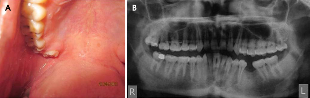
Fig. 2 Case 2. A. Intraoral photograph shows a supernumerary tooth, placed palatal and distal to the third molar. B. Panoramic radiograph shows a supernumerary tooth distal to the right maxillary third molar.
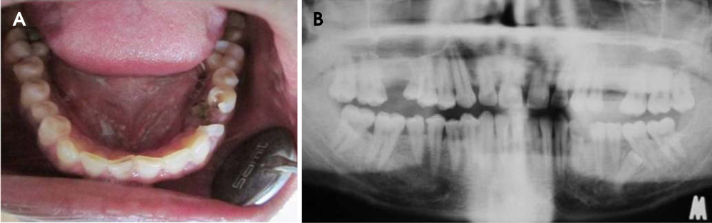
Fig. 3 Case 3. A. Intraoral photograph shows a carious lesion of the left mandibular first premolar, with the presence of root piece of the second premolar followed by two more premolar-like teeth in the lower left quadrant. On the right side, there are a total of 4 well aligned premolars. B. Panoramic radiograph shows a supernumerary tooth distal to the right maxillary premolars, two erupted supernumerary teeth on the right mandibular premolar area, and an impacted supernumerary tooth mesial to the left mandibular first molar.

Fig. 4 Case 4. A. Intraoral photograph shows three supernumerary teeth lingual and distal to the right mandibular premolars. B. Panoramic radiograph reveals an impacted supplementary tooth apical to the left mandibular second premolar. C. Intraoral periapical radiograph shows the presence of a dilacerated premolar-like tooth between the second premolar and the first molar.
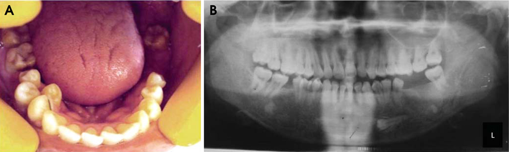
Fig. 5 Case 5. A. Intraoral photograph shows a supplementary tooth distal to the left mandibular premolar and two supplementary teeth lingual and distal to the right mandibular premolars. B. Panoramic radiograph reveals an impacted supplementary tooth below the left mandibular canine and premolars.
Reference
-
1. Leco Berrocal MI, Martín Morales JF, Martínez González JM. An observational study of the frequency of supernumerary teeth in a population of 2000 patients. Med Oral Patol Oral Cir Bucal. 2007. 12:E134–E138.2. Hurlen B, Humerfelt D. Characteristics of premaxillary hyperodontia. A radiographic study. Acta Odontol Scand. 1985. 43:75–81.
Article3. Tommasi AF. Diagnostico em patologia bucal. 1988. Sao Paulo: Artes Medicas;85–97.4. Rao PV, Chidzonga MM. Supernumerary teeth: literature review. Cent Afr J Med. 2001. 47:22–26.
Article5. Batra P, Duggal R, Parkash H. Non-syndromic multiple supernumerary teeth transmitted as an autosomal dominant trait. J Oral Pathol Med. 2005. 34:621–625.
Article6. Sedano HO, Gorlin RJ. Familial occurrence of mesiodens. Oral Surg Oral Med Oral Pathol. 1969. 27:360–361.
Article7. Yusof WZ. Non-syndrome multiple supernumerary teeth: literature review. J Can Dent Assoc. 1990. 56:147–149.8. Garvey MT, Barry HJ, Blake M. Supernumerary teeth - an overview of classification, diagnosis and management. J Can Dent Assoc. 1999. 65:612–616.9. Giancotti A, Grazzini F, De Dominicis F, Romanini G, Arcuri C. Multidisciplinary evaluation and clinical management of mesiodens. J Clin Pediatr Dent. 2002. 26:233–237.10. Mishra MB. Types of hyperodontic anomalies in permanent dentition: report of 5 cases. Int J Clin Dent Sci. 2011. 2:15–21.11. Hegde SV, Munshi AK. Late development of supernumerary teeth in the premolar region: a case report. Quintessence Int. 1996. 27:479–481.12. Mason C, Rule DC, Hopper C. Multiple supernumeraries: the importance of clinical and radiographic follow-up. Dentomaxillofac Radiol. 1996. 25:109–113.
Article
- Full Text Links
-
- Actions
-
Cited
- CITED
-
- Close
- Share
-
- Similar articles
-
- BBilateral Intranasal Supernumerary Teeth
- A Case of Supernumerary Tooth in Tonsil
- Prevalence of Dental Anomalies in Patients with Non-syndromic Cleft Lip with or without Cleft Palate
- Two Cases of Mesiodens in Nasal Cavity
- Immediate placement of implant following extraction of impacted supernumerary teeth and permanent teeth : A case report
