Septic Hip Arthritis with Iliopsoas Abscess Detected after Spine Operation: A Case Report
- Affiliations
-
- 1Department of Orthopaedic Surgery, Pusan National University School of Medicine, Busan, Korea. cibacoll@hanmail.net
- KMID: 1974381
- DOI: http://doi.org/10.5371/hp.2013.25.3.237
Abstract
- In the elderly patients who complain of pain in the buttock and leg, it is not easy to distinguish whether the pain comes from the lesion of the hip or from the spine. A 78-year-old female who was treated conservatively for persistent pain in the right buttock and leg after an operation for spinal stenosis in the local clinic visited our clinic. Septic hip arthritis with severe femoral head destruction and multiple abscesses in the buttock and iliopsoas muscle were diagnosed 2 months postoperatively, and spinal abscess in the site of the previous operation was detected by a subsequent MRI study. To avoid such a delay of the diagnosis and treatment, it is important to suspect hip joint lesion earlier for the source of persistent pain after a spine operation. Further more, diagnostic evaluation is necessary to rule out co-infection of the spine or iliopsoas muscle when a hip joint infection exists.
MeSH Terms
Figure
-
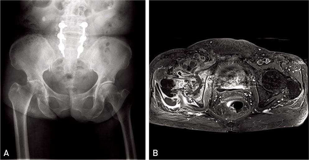
Fig. 1 (A) Plain X-ray shows femoral head destruction with acetabuluar subchondral bone involvement in the right hip joint. (B) MRI shows septic arthritis and buttock abscess with psoas muscle involvement in the right hip joint.

Fig. 2 (A) MRI shows right iliopsoas muscle infection. (B) MRI shows infectious finding in the previous spine operation site.
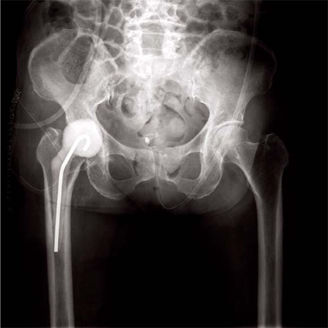
Fig. 3 Plain X-ray shows insertion state of unipolar antibiotics-mixed cement spacer after femoral head resection and debridement of right hip joint.
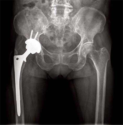
Fig. 4 Plain X-ray shows the last follow up after the revision of 2nd stage reimplantation.
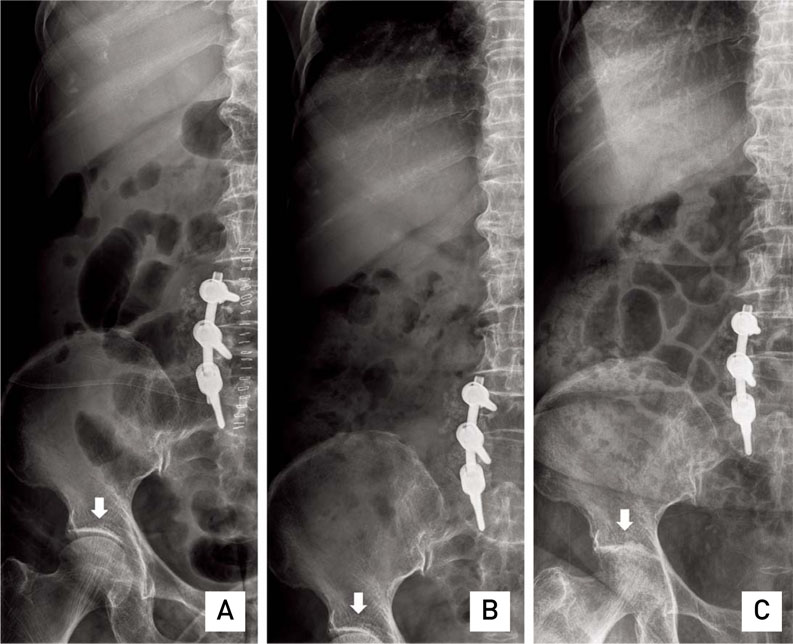
Fig. 5 Serial follow up lumbar plain X-rays checked at postoperative (A) 1 day, (B) 4 weeks, and (C) 8 weeks show gradual narrowing and destruction of the hip joint space in the right hip.
Reference
-
1. Kumagai K, Ushiyama T, Kawasaki T, Matsusue Y. Extension of lumbar spine infection into osteoarthritic hip through psoas abscess. J Orthop Sci. 2005; 10:91–94.
Article2. Gavet F, Tournadre A, Soubrier M, Ristori JM, Dubost JJ. Septic arthritis in patients aged 80 and older: a comparison with younger adults. J Am Geriatr Soc. 2005; 53:1210–1213.
Article3. Gruenwald I, Abrahamson J, Cohen O. Psoas abscess: case report and review of the literature. J Urol. 1992; 147:1624–1626.
Article4. Molloy A, Laing A, O'Shea K, Bell L, O'Rourke K. The complications of septic arthritis in the elderly. Aging Clin Exp Res. 2010; 22:270–273.
Article5. Dala-Ali BM, Lloyd MA, Janipireddy SB, Atkinson HD. A case report of a septic hip secondary to a psoas abscess. J Orthop Surg Res. 2010; 5:70.
Article6. Sadat-Ali M, al-Habdan I, Ahlberg A. Retrofascial nontuberculous psoas abscess. Int Orthop. 1995; 19:323–326.
Article
- Full Text Links
-
- Actions
-
Cited
- CITED
-
- Close
- Share
-
- Similar articles
-
- An Enlarged Iliopsoas Bursa Associated with Osteonecrosis of the Femoral Head: A Case Report
- Rapid Destruction of the Hip Joint Accompanied by an Enlarged Iliopsoas Bursa in a Healthy Man
- Arthroscopic Treatment of Septic Arthritis of the Hip in a Child: A Case Report
- Femoral Nerve Palsy caused by Iliopsoas Bursitis Associated with Osteonecrosis of the Femoral Head : A Case Report
- Mycoplasma hominis Septic Arthritis of the Hip Developed in the Postpartum Period
