Two Cases of the Diffuse Sclerosing Variant of Papillary Thyroid Carcinoma
- Affiliations
-
- 1Department of Internal Medicine, Konyang University Medical School, Daejeon, Korea.
- 2Department of Pathology, Konyang University Medical School, Daejeon, Korea.
- KMID: 1965997
- DOI: http://doi.org/10.3803/jkes.2008.23.6.430
Abstract
- The diffuse sclerosing variant of papillary thyroid carcinoma (DSPTC) is a rare histological subtype characterized by diffuse involvement of one or both thyroid lobes, widespread lymphatic permeation, prominent fibrosis, squamous metaplasia, abundant psammoma body and lymphatic infiltration. This subtype usually occurs in young female, and exhibits a higher frequency of cervical and distant metastasis. DSPTC clinically resembles Hashimoto's thyroiditis, and often delays the correct diagnosis. We experienced two patients with DSPTC: the one patient presented with a neck mass lasting for a month, and in the other patient, a thyroid lesion was incidentally found during a medical examination.
MeSH Terms
Figure
-
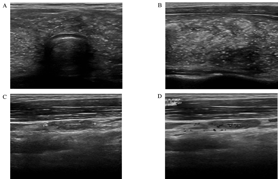
Fig. 1 Ultrasonographic finding of neck shows diffuse enlargement of thyroid gland with diffuse scattered tiny hyperechogenic spots and multinodular appearance in entire thyroid gland (A, B), and small sized lymphadenopathy with abnormal echogenecity in both thyroid (C, D).
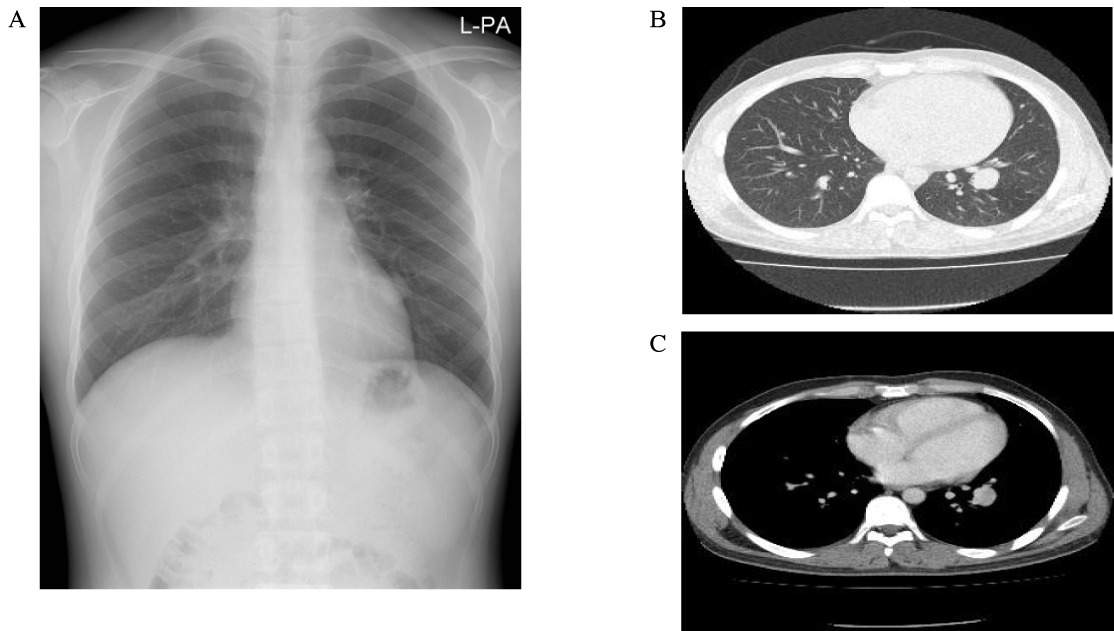
Fig. 2 Simple chest X-ray findings of the patient shows about 2.1 cm-sized, well marginated nodule in left lower lobe (A). Chest CT shows about 2.1 cm-sized, well marginated heterogenous enhancing nodule in left lower lobe suggesting benign mass such as sclerosing hemangioma or inflammatory granuloma rather than metastasis (B, C).
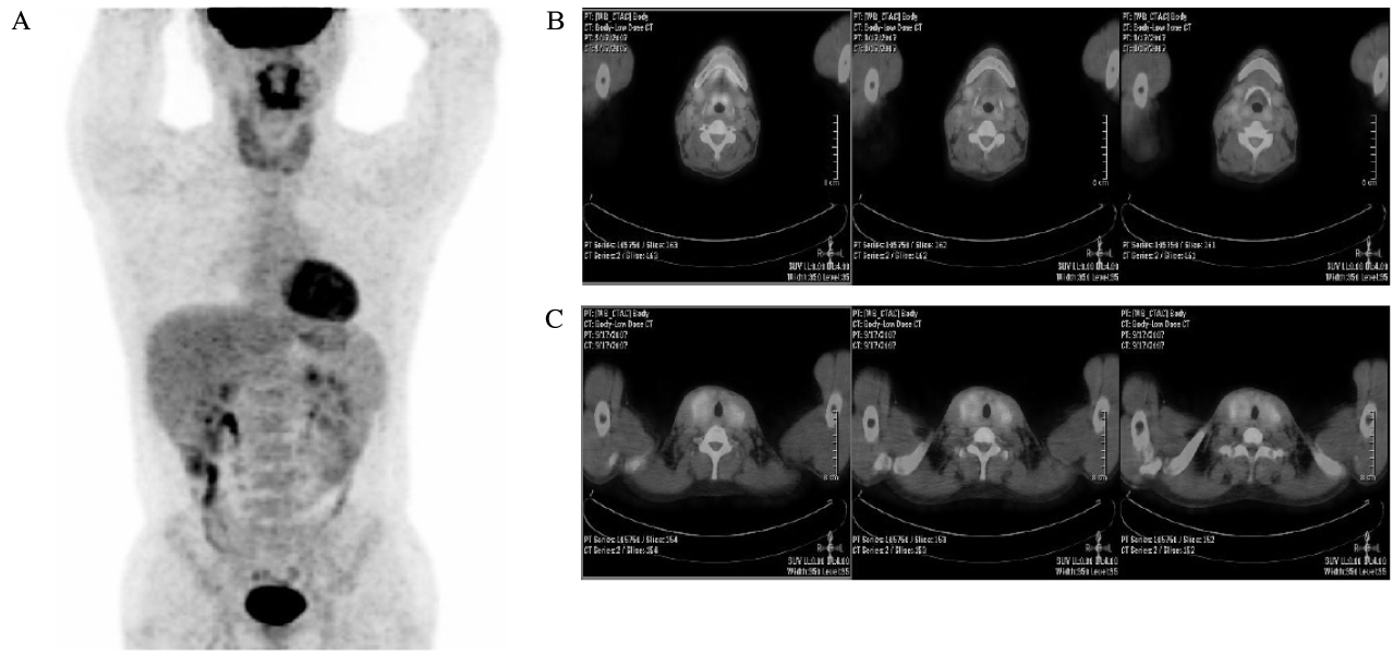
Fig. 3 PET-CT scan shows diffuse increased FDG uptake in thyroid gland (A), multiple nodal activities in the right internal jugular chain (B, C), and no other abnormal FDG uptake in the body (A).
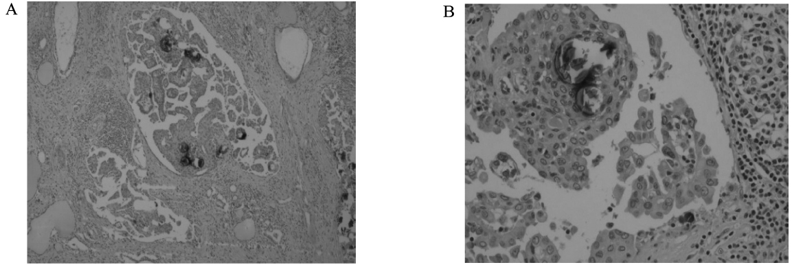
Fig. 4 Microscopic finding: Tumor has multiple nodular structures in the entire thyroidal parenchyma and shows lymphocytic infiltration and fibrosis (A). Nodular structures consist of typical tumor cells of papillary thyroid carcinoma with nuclear groove and intranuclear inclusions. Psammoma bodies and squamous metaplasia are frequently observed (B). (H&E stain, ×40 (A), ×200 (B))
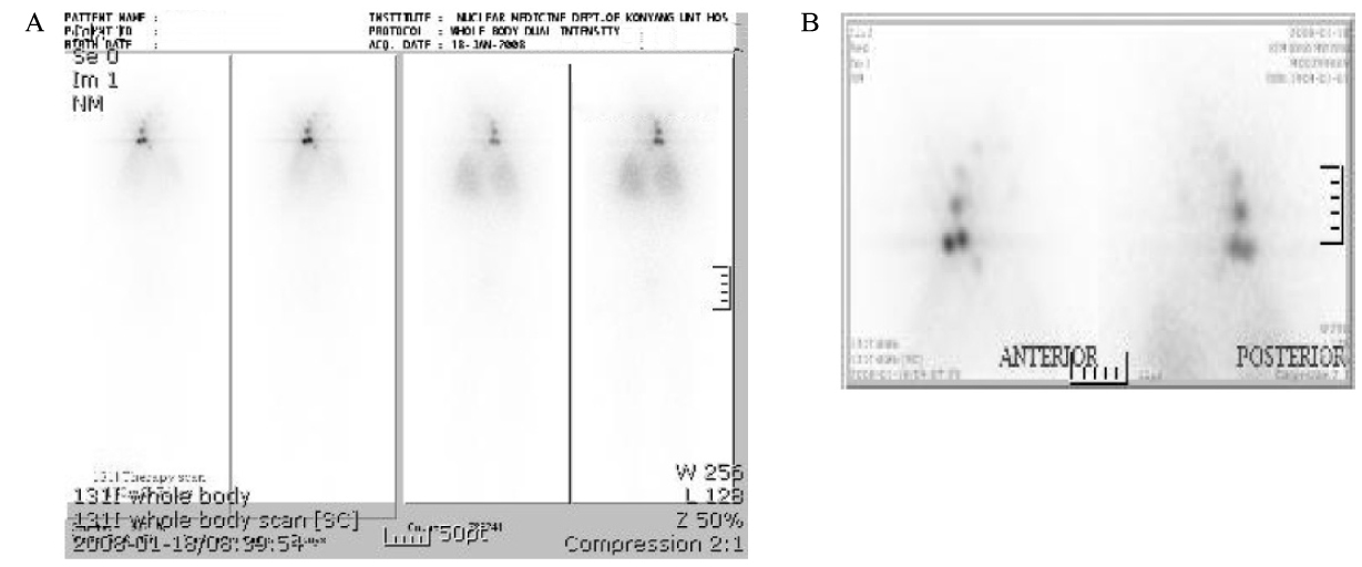
Fig. 5 Post-therapy scan shows multiple increased uptakes in both anterior neck, suggesting metastatic lymphadenopathy. There is no evidence of distant metastasis (A, B).

Fig. 6 Ultrasonographic finding of neck shows heterogenous parenchymal echo with ill-defined hyper- and hypoechoic lesions in right lobe of thyroid (A, B), and multiple abnormal enlarged lymph nodes with/without intranodal calcification, abnormal vascularity, and some cystic changes in both lobe of thyroid (C, D).

Fig. 7 Neck CT of the patient shows about 9 mm size, ill-defined low density nodule in right lobe of thyroid gland (A), and enlarged lymph node suggesting metastasis in right level IV of neck (B).

Fig. 8 Microscopic finding: Thyroid gland shows multiple variable-sized nodular structures with numerous psammoma bodies, lymphocytic infiltration and fibrosis in entire thyroid parenchyma (A). Nodular structures show typical tumor cells of papillary thyroid carcinoma with intranuclear inclusion and multiple squamous metaplasia (B). (H&E stain, ×40 (A), ×200 (B))
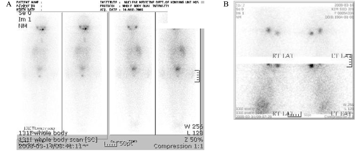
Fig. 9 Posttheraty scan shows no abnormal uptake (A, B).
Reference
-
1. Crile G Jr, Fisher ER. Simultaneous occurrence of thyroiditis and papillary carcinoma: report of two cases. Cancer. 1953. 6:57–62.2. Fujimoto Y, Obara T, Ito Y, Kodama T, Aiba M, Yamaguchi K. Diffuse sclerosing variant of papillary carcinoma of the thyroid. clinical importance, surgical treatment, and follow-up study. Cancer. 1990. 66:2306–2312.3. Lee SH, Lee YS, Yun JS, Jeong JJ, Nam KH, Chung WY, Chang HS, Park JS. Diffuse sclerosing variant of papillary thyroid carcinoma: a 17-year experience at a single institution. J Korean Surg Soc. 2008. 74:98–104.4. Soares J, Limbert E, Sobrinho-Simoes M. Diffuse sclerosing variant of papillary thyroid carcinoma: a clinicopathologic study of 10 cases. Pathol Res Pract. 1989. 185:200–206.5. Schlumberger M, Pacini F. Thyroid tumors. 2006. 3rd ed. Paris: Nucleon;42–43.6. Vickery AL Jr, Carcangiu ML, Johannessen JV, Sobrinho-Simoes M. Papillary carcinoma. Semin Diagn Pathol. 1985. 2:90–100.7. Wu PS, Leslie PJ, McLaren KM, Toft AD. Diffuse sclerosing papillary carcinoma of thyroid: a wolf in sheep's clothing. Clin Endocrinol. 1989. 31:535–540.8. Carcangju ML, Bianchi S. Diffuse sclerosing variant of papillary thyroid carcinoma: clinicopathologic study of 15 cases. Am J Surg Pathol. 1989. 13:1041–1049.9. Albareda M, Puig-Domingo M, Wengrowicz S, Soldevila J, Matias-Guju X, Caballero A, Chico A, De Leiva A. Clinical forms of presentation and evolution of diffuse sclerosing variant of papillary carcinoma and insular variant of follicular carcinoma of the thyroid. Thyroid. 1998. 8:385–391.10. Lam AK, Lo CY. Diffuse sclerosing variant of papillary carcinoma of thyroid: a 35-year comparative study at a single institution. Ann Surg Oncol. 2006. 13:176–181.11. Kobayashi K, Fukata S, Amino N, Miyauchi A. A case with diffuse sclerosing variant of papillary carcinoma of the thyroid: characteristic features on ultrasonography. J Med Ultrasonics. 2006. 33:159–161.12. Chow SM, Chan JK, Law SC, Tang DL, Ho CM, Cheung WY, Wong IS, Lau WH. Diffuse sclerosing variant of papillary thyroid carcinoma-clinical features and outcome. Eur J Surg Oncol. 2003. 29:446–449.13. Imamura Y, Kasahara Y, Fukuda M. Multiple brain metastases from a diffuse sclerosing variant of papillary carcinoma of the thyroid. Endocr Pathol. 2000. 11:97–108.14. Falvo L, Giacomelli L, D'Andrea V, Marzullo A, Guerriero G, de Antoni E. Prognostic importance of sclerosing variant in papillary thyroid carcinoma. Am Surg. 2006. 72:438–444.15. Thompson LD, Wieneke JA, Heffess CS. Diffuse sclerosing variant of papillary thyroid carcinoma: a clinicopathologic and imunophenotypic analysis of 22 cases. Endocr Pathol. 2005. 16:331–348.16. Kwak JY, Kim EK, Hong SW, Oh KK, Kim MJ, Park CS, Cheong WY. Diffuse sclerosing variant of papillary carcinoma of the thyroid: ultrasound features with histopathological correlation. Clin Radiol. 2007. 62:382–386.17. Lee SC, Kim DW. Diffuse sclerosing variant of papillary thyroid carcinoma: case report. J Korean Radiol Soc. 2006. 55:49–52.18. Ohori NP, Schoedel KE. Cytopathology of high grade papillary thyroid carcinomas: tall-cell variant, diffuse sclerosing variant, and poorly differentiated papillary carcinoma. Diagn Cytopathol. 1999. 20:19–23.19. Kim JM, Kim MR, Min SK, Chu YC, Kim KR. Fine needle aspiration cytology of diffuse sclerosing variant of papillary carcinoma of the thyroid: case report. Korean J Cytopathol. 2000. 11:47–52.20. Chan JK, Tsui MS, Tse CH. Diffuse sclerosing variant of papillary carcinoma of the thyroid. A histological and immunohistochemistry of three cases. Histopathology. 1987. 11:191–201.
- Full Text Links
-
- Actions
-
Cited
- CITED
-
- Close
- Share
-
- Similar articles
-
- Diffuse Sclerosing Variant of Papillary Thyroid Carcinoma: Case Report
- Papillary Thyroid Carcinoma of a Diffuse Sclerosing Variant: Ultrasonographic Monitoring from a Normal Thyroid Gland to Mass Formation
- Diffuse Sclerosing Variant of Papillary Thyroid Carcinoma in a Child: A Case Report
- Fine Needle Aspiration Cytology of Diffuse Sclerosing Variant of Papillary Carcinoma of the Thyroid: A Case Report
- A Case Report on Diffuse Sclerosing Papillary Carcinoma of the Thyroid: The Ultrasound and CT Images
