Introduction to Starting Upper Gastrointestinal Endoscopy: Proper Insertion, Complete Observation, and Appropriate Photographing
- Affiliations
-
- 1Department of Internal Medicine, Keimyung University School of Medicine, Daegu, Korea. seenae99@dsmc.or.kr
- KMID: 1964268
- DOI: http://doi.org/10.5946/ce.2015.48.4.279
Abstract
- Diagnostic upper gastrointestinal endoscopy is the most basic of endoscopy procedures and is the technique that trainee doctors first learn. Mastering the basics of endoscopy is very important because when this process is imprecise or performed incorrectly, it can severely affect a patient's health or life. Although there are several guidelines and studies that consider these basics, there are still no standard recommendations for endoscopy in Korea. In this review, basic points, including proper endoscope insertion, precise observation without blind spots, and appropriate photographing, for upper gastrointestinal endoscopy will be discussed.
Figure
-
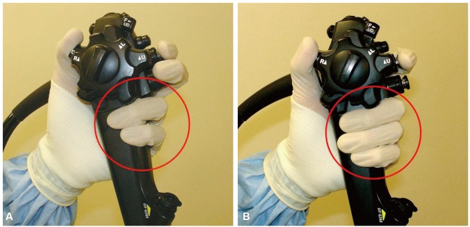
Fig. 1 Grips in the endoscopic procedure. (A) Two-finger method, in which the ring and little fingers hold the endoscope and the index and middle fingers operate the suction and aspiration valves. (B) Three-finger method, in which the middle, ring, and little fingers hold the endoscope and only the index finger is used for the suction and aspiration valves. Adapted from Lee,13 with permission from EndoTODAY.
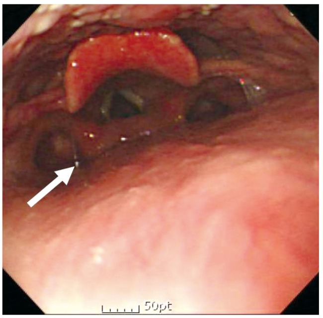
Fig. 2 When the endoscope arrives at the left pyriform sinus (arrow) as seen on the left side of the screen, its tip is maneuvered so that it faces the center and the endoscope is carefully inserted.
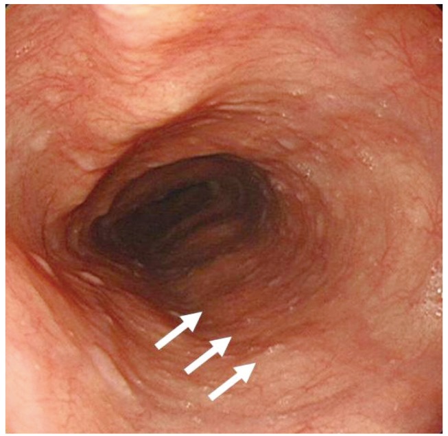
Fig. 3 Compression from the spine (arrows). Care must be taken not to mistake these for pathological lesions.
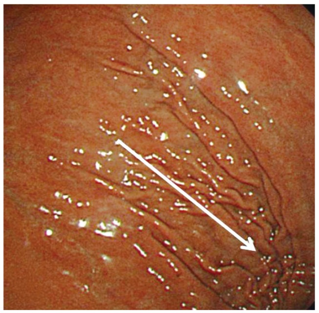
Fig. 4 As the tube enters the stomach, gastric folds can be observed in the 10 and 4 o'clock positions.
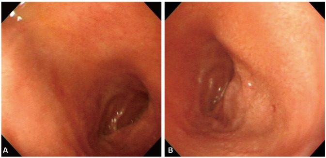
Fig. 5 Duodenal bulb. (A) The anterior wall and the lesser curvature can be observed simultaneously in the 8 and 11 o'clock positions, respectively. (B) As the left arm is rotated clockwise, the posterior wall and greater curvature are observed simultaneously in the 2 and 5 o'clock positions.
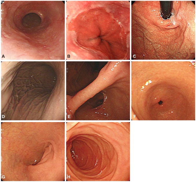
Fig. 6 Essential photographing sites. (A) Upper esophagus. (B) Lower esophagus. (C) Gastric cardia and fundus. (D) Upper body. (E) Gastric angle. (F) Gastric antrum. (G) Duodenal bulb. (H) Duodenal second portion.
Cited by 1 articles
-
Highlights from the 52nd Seminar of the Korean Society of Gastrointestinal Endoscopy
Eun Young Kim, Il Ju Choi, Kwang An Kwon, Ji Kon Ryu, Ki Baik Hahm
Clin Endosc. 2015;48(4):269-278. doi: 10.5946/ce.2015.48.4.269.
Reference
-
1. Cappell MS. Safe "hands-on" teaching of endoscopy to beginning gastroenterology fellows. Gastrointest Endosc. 2011; 73:847. PMID: 21457821.
Article2. Northup PG, Argo CK, Muir AJ, Decross AJ, Coyle WJ, Oxentenko AS. Procedural competency of gastroenterology trainees: from apprenticeship to milestones. Gastroenterology. 2013; 144:677–680. PMID: 23439111.
Article3. Multisociety Task Force on GI Training. Report of the multisociety task force on GI training. Am J Gastroenterol. 2009; 104:2659–2663. PMID: 19888229.4. Cha JM. Quality improvement of gastrointestinal endoscopy in Korea: past, present, and future. Korean J Gastroenterol. 2014; 64:320–332. PMID: 25530583.
Article5. Kwon KA, Choi IJ, Kim EY, Dong SH, Hahm KB. Highlights of the 48th seminar of korean society of gastrointestinal endoscopy. Clin Endosc. 2013; 46:203–211. PMID: 23767027.
Article6. Lee YK, Park JB. Steps of reprocessing and equipments. Clin Endosc. 2013; 46:274–279. PMID: 23767039.
Article7. Park JM. Quality control for upper gastrointestinal endoscopy. Korean J Gastrointest Endosc. 2010; 40:343–346.8. Armstrong D, Barkun A, Bridges R, et al. Canadian Association of Gastroenterology consensus guidelines on safety and quality indicators in endoscopy. Can J Gastroenterol. 2012; 26:17–31. PMID: 22308578.
Article9. ASGE Standards of Practice Committee. Jain R, Ikenberry SO, et al. Minimum staffing requirements for the performance of GI endoscopy. Gastrointest Endosc. 2010; 72:469–470. PMID: 20579993.
Article10. Aabakken L, Rembacken B, LeMoine O, et al. Minimal standard terminology for gastrointestinal endoscopy: MST 3.0. Endoscopy. 2009; 41:727–728. PMID: 19670144.11. Rey JF, Lambert R. ESGE Quality Assurance Committee. ESGE recommendations for quality control in gastrointestinal endoscopy: guidelines for image documentation in upper and lower GI endoscopy. Endoscopy. 2001; 33:901–903. PMID: 11605605.
Article12. Standards of practice of gastrointestinal endoscopy. Guidelines. Gastrointest Endosc. 1988; 34(3 Suppl):8S. PMID: 3384294.13. Lee JH. Inserting endoscope [Internet]. Seoul: EndoTODAY;2013. cited 2015 Jun 10. Available at: http://endotoday.com.
- Full Text Links
-
- Actions
-
Cited
- CITED
-
- Close
- Share
-
- Similar articles
-
- Upper gastrointestinal diseases diagnosed by upper gastrointestinal fiberoptic endoscopy in children
- Observable Laryngopharyngeal Lesions during the Upper Gastrointestinal Endoscopy
- Observation Time for Complete Endoscopy in Gastric Cancer Screening
- Compromised ventilation caused by tracheoesophageal fistula and gastrointestinal endoscope undergoing removal of disk battery on esophagus in pediatric patient -A case report-
- Management of aerosol generation during upper gastrointestinal endoscopy
