Determination of the Posture of Transplanted Ear Region Ectoderm in Amphibian Embryoes
- Affiliations
-
- 1Department of Anatomy, Severance Union Medical College, Seoul, Korea.
- KMID: 1963574
- DOI: http://doi.org/10.3349/JSeveranceMed.1933.1.1.5
Abstract
- No abstract available. [EXPERIMENTAL METHOD AND MATERIALS: All the experiments were carried out on the embryo of Rana temporaria at two stages as follows; 1. The FIRST STAGE is that of the earliest appearance of the neural folds. The ectoderm of the ear region consists of two layer of epithelial cells and no difference from the neighbouring ectoderm is apparent. 2. SECOND STAGE is that of closure of the neural folds. The ectoderm of the ear region is slightly thickened as a few layers of epithelial cells and is thus differentiated from the neighbouring ectoderm. Operative work was done in 0.2% saline solution, under the binocular microscope using fine needles for dissection. A piece of ectoderm 0.5 mm square was removed from the left ear region and replaced after rotating 180 degrees. The anterior part of the piece comes to lie posteriorly and the superior part of the piece comes to lie at the lowest part of the graft. The specimens were allowed to go on with their development for from 42 to 45 days, at the end of which time they were preserved in bichloride-picric acid solution. The preserved specimens were embedded in paraffin, cut in serial sections and stained with eosin-haematoxylin. Reconstruction wax-plate models after Born's method were made. The ear region of each specimen examined as to form, position, posture, size, semicircular canals, endolymphatic appendage, lagena, accompanying nerves and ganglia and the cartilaginous capsule.]
MeSH Terms
Figure
-

Fig. No. I Wax-plate model, Op. No. I, 6-8. 100 x. Operation after 45 days. Transplantation of the ectoderm of left ear region rotated to 180 degrees. The position of the ear was little changed but the ear is normal left side ear. (FIRST STAGE)
Fig. No. II Cross section of the specimen of Op. No. I, 6-8. 82 x.
Fig. No. III Wax-plate model, Op. No. II, 7-4. 100 x. Ope- Cross section of specimen of ration after 45 davs. Transplantation of the ecto- Op. No. II, 7-4. 82 x. derm of left ear region rotated to 180 degrees. Its posture was reversed antero-posteriorly, and superio- inferiorly.
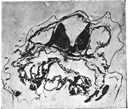
Fig. No. IV Cross section of the specimen of Op. No. II, 7-4. 82 x.
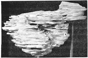
Fig. No. V Wax-plate model, Op. No. IV, 2-7. 100 x. Cross section of specimen of Op. Operation after 42 days. Transplantation of the No. IV, 2-7. 82 x ectoderm of left side ear region to the middle part of the lateral line after rotation through 180 degrees. The ear is reversed antero-posteriorly.
Fig. No. VI Cross section of the specimen of Op. No. IV, 2-7. 82 x.
- Full Text Links
-
- Actions
-
Cited
- CITED
-
- Close
- Share
-
- Similar articles
-
- A Case of Teratoma of the Middle Ear
- A Case of Postauricular Dermoid Cyst
- Reproducibility and reliability of head posture obtained by the outer canthus indicator
- Transition from Canalolithiasis to Cupulolithiasis by the Head-Bending Posture and Canalith Repositioning by Using the Side-Lying Position in Benign Paroxysmal Positional Vertigo of Horizontal Semicircular Canal
- Congenital Dermal Sinus at Thoracic Region Associated
