Verruciform xanthoma of the palatal gingiva: a report of two cases
- Affiliations
-
- 1Department of Oral and Maxillofacial Surgery, Gangnam Severance Hospital, College of Dentistry, Yonsei University, Seoul, Korea. omspark@yuhs.ac
- 2Department of Oral Pathology, Oral Cancer Research Institute, College of Dentistry, Yonsei University, Seoul, Korea.
- 3Department of Oral and Maxillofacial Surgery, College of Dentistry, Yonsei University, Seoul, Korea.
- KMID: 1960980
- DOI: http://doi.org/10.5125/jkaoms.2013.39.6.292
Abstract
- Verruciform xanthoma (VX) is a rare, benign lesion that presents in the oral cavity, skin, or genital organs as a verrucous, papillomatous, or flat papule with varying colors. VX has indistinct clinical features, making histopathological examination necessary for a definitive diagnosis. Histologically, VX is characterized by parakeratosis, rete ridges with uniform depth, and an accumulation of the foam cells, which are also known as the "xanthoma cells". These foam cells test positive for antibodies, such as CD-68 and vimentin; it is thought that VX foam cells are derived from the monocyte-macrophage lineage, and that VX's pathogenic mechanism is partly related to an immune mechanism. Nevertheless, the pathogenesis of VX remains unclear. VX can be treated by surgical excision; other medical, chemical, and radiological treatments are not required postoperatively. Recurrence and malignant transformation of VX are rare. Two patients, each with a mass of unknown origin on the palatal gingiva, were presented at our clinic. Excisional biopsies of the masses were performed for a histological diagnosis after clinical and radiological examinations. Histological examination confirmed a diagnosis of VX in both cases.
Keyword
MeSH Terms
Figure
-
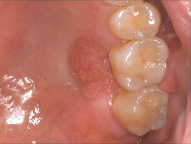
Fig. 1 Clinical aspect of the palate lesion in Case 1 on the first visit. A reddish papule with a rough verrucous surface and distinctive margin can be observed.
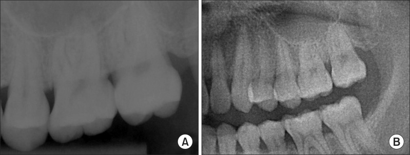
Fig. 2 Radiological examination of Case 1. A. Periapical view. B. A magnified view of orthopantogram. No specifing findings on these two radiograph images.
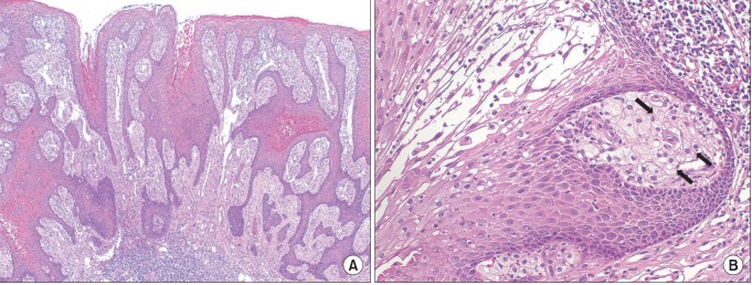
Fig. 3 Histopathological examination of Case 1 after excisional biopsy. A. The tissue has a papillary appearance with hyperparakeratosis and uniform rete peg (H&E staining, ×40). B. High magnification (H&E staining, ×200) illustrates large foam cells (xanthoma cells, black arrows) in the connective tissue papillae.
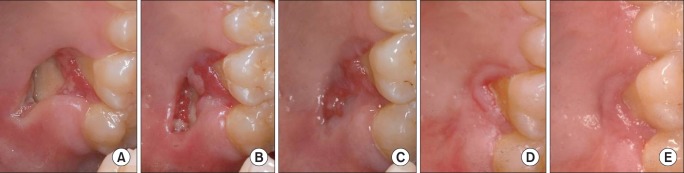
Fig. 4 Follow-up clinical photos of Case 1 representing a good secondary healing state. A. Two weeks postoperative. B. Three weeks postoperative. C. Five weeks postoperative. D. Four months postoperative. E. One year postoperative.

Fig. 5 Histopathological examination of Case 2 at the diagnostic incisional biopsy. A. Papillary surface with parakeratosis (H&E staining, ×100). B. Xanthoma cells (black arrows) (H&E staining, ×400).
Cited by 1 articles
-
Verruciform xanthoma in the hard palate: a case report and literature review
Alexandre Simões Garcia, Otávio Pagin, Paulo Sérgio da Silva Santos, Denise Tostes Oliveira
J Korean Assoc Oral Maxillofac Surg. 2016;42(6):383-387. doi: 10.5125/jkaoms.2016.42.6.383.
Reference
-
1. Shafer WG. Verruciform xanthoma. Oral Surg Oral Med Oral Pathol. 1971; 31:784–789. PMID: 5280461.
Article2. Zegarelli DJ, Zegarelli-Schmidt EC, Zegarelli EV. Verruciform xanthoma. Further light and electron microscopic studies, with the addition of a third case. Oral Surg Oral Med Oral Pathol. 1975; 40:246–256. PMID: 1057149.3. van der Waal I, Kerstens HC, Hens CJ. Verruciform xanthoma of the oral mucosa. J Oral Maxillofac Surg. 1985; 43:623–626. PMID: 3859615.
Article4. Philipsen HP, Reichart PA, Takata T, Ogawa I. Verruciform xanthoma--biological profile of 282 oral lesions based on a literature survey with nine new cases from Japan. Oral Oncol. 2003; 39:325–336. PMID: 12676251.
Article5. Yu CH, Tsai TC, Wang JT, Liu BY, Wang YP, Sun A, et al. Oral verruciform xanthoma: a clinicopathologic study of 15 cases. J Formos Med Assoc. 2007; 106:141–147. PMID: 17339158.
Article6. Nam YH, Oh CW. A case of verruciform xanthoma presenting as leukoplakia on the lower lip. Korean J Dermatol. 2006; 44:508–511.7. Choi YH, Moon YJ, Lee YW, Won JY, Song ES. A case of multiple verruciform xanthoma of oral cavity and gastrointestinal tract. Korean J Dermatol. 2002; 40:162–165.8. Travis WD, Davis GE, Tsokos M, Lebovics R, Merrick HF, Miller SP, et al. Multifocal verruciform xanthoma of the upper aerodigestive tract in a child with a systemic lipid storage disease. Am J Surg Pathol. 1989; 13:309–316. PMID: 2539022.
Article9. Mete O, Kurklu E, Bilgic B, Beka H, Unur M. Flat-type verruciform xanthoma of the tongue and its differential diagnosis. Dermatol Online J. 2009; 15:5. PMID: 19930992.
Article10. Neville BW, Damm DD, Allen CM, Bouquot JE. Oral & maxillofacial pathology. 2nd ed. Philadelphia: WB Saunders;2002.11. Cumberland L, Dana A, Resh B, Fitzpatrick J, Goldenberg G. Verruciform xanthoma in the setting of cutaneous trauma and chronic inflammation: report of a patient and a brief review of the literature. J Cutan Pathol. 2010; 37:895–900. PMID: 19958440.
Article12. Shahrabi Farahani S, Treister NS, Khan Z, Woo SB. Oral verruciform xanthoma associated with chronic graft-versus-host disease: a report of five cases and a review of the literature. Head Neck Pathol. 2011; 5:193–198. PMID: 21305367.13. Hu JA, Li Y, Li S. Verruciform xanthoma of the oral cavity: clinicopathological study relating to pathogenesis. Report of three cases. APMIS. 2005; 113:629–634. PMID: 16218939.
Article14. Mostafa KA, Takata T, Ogawa I, Ijuhin N, Nikai H. Verruciform xanthoma of the oral mucosa: a clinicopathological study with immunohistochemical findings relating to pathogenesis. Virchows Arch A Pathol Anat Histopathol. 1993; 423:243–248. PMID: 8236821.
Article15. Visintini E, Rizzardi C, Chiandussi S, Biasotto M, Melato M, Di Lenarda R. Verruciform xanthoma of the oral mucosa. Report of a case. Minerva Stomatol. 2006; 55:639–645. PMID: 17211369.
- Full Text Links
-
- Actions
-
Cited
- CITED
-
- Close
- Share
-
- Similar articles
-
- A Case of Verruciform Genital-associated (Vegas) Xanthoma on the Scrotum
- Clinical, histological, and deep learning-based assessments and treatment of oral verruciform xanthoma: a case report
- A Case of Verruciform Xanthoma on the Scrotum
- A Case of Verruciform Xanthoma Associated with Epidermal Atypia
- Verruciform Xanthoma of the Penis
