J Korean Assoc Oral Maxillofac Surg.
2013 Dec;39(6):263-268. 10.5125/jkaoms.2013.39.6.263.
Distribution of the lingual foramina in mandibular cortical bone in Koreans
- Affiliations
-
- 1Department of Oral and Maxillofacial Surgery, College of Dentistry, Dankook University, Cheonan, Korea. kimchoms@dankook.ac.kr
- KMID: 1960975
- DOI: http://doi.org/10.5125/jkaoms.2013.39.6.263
Abstract
OBJECTIVES
The interforminal region, between the mandibular foramen, is known as a relatively safe area that is free of anatomic structures, such as inferior alveolar nerve, submandibular fossa, and lingual side of the mandible is occasionally neglected for its low clinical importance. Even in the case of a severely constricted alveolus, perforation of the lingual cortical bone had been intended. However, anterior extension of the inferior alveolar canal, important anatomic structure, such as concavity of lingual bone, lingual foramina, and lingual canal, has recently been reported through various studies, and untypical bleeding by perforation of the lingual plate on implantation has also been reported. Therefore, in this study, we performed radiographic and statistical analysis on distribution and appearance frequencies of the lingual foramina that causes perforation of the mandibular lingual cortical bone to prevent complications, such as untypical bleeding, during surgical procedure.
MATERIALS AND METHODS
We measured the horizontal length from a midline of the mandible to the lingual foramina, as well as the horizontal length from the alveolar crest to the lingual foramina and from the lingual foramina to the mandibular border by multi-detector computed tomography of 187 patients, who visited Dankook University Dental Hospital for various reasons from January 1, 2008 to August 31, 2012.
RESULTS
From a total of 187 human mandibles, 110 (58.8%) mandibles had lingual foramina; 39 (20.9%) had bilateral lingual foramen; 34 (18.2%) had the only left lingual foramen; and 37 (19.8%) had the only right lingual foramen.
CONCLUSION
When there is consistent bleeding during a surgical procedure, clinicians must consider damages on the branches of the sublingual artery, which penetrate the lingual foramina. Also, when there is a lingual foramina larger than 1 mm in diameter on a pre-implantation computed tomography, clinicians must beware of vessel damage. In order to prevent these complications and progress with a safe surgical procedure, a thorough radiographic examination before the surgery is indispensable. Further, clinicians should retract lingual flap definitely to confirm the shape of the lingual bone and existence of the lingual foramina.
MeSH Terms
Figure
-
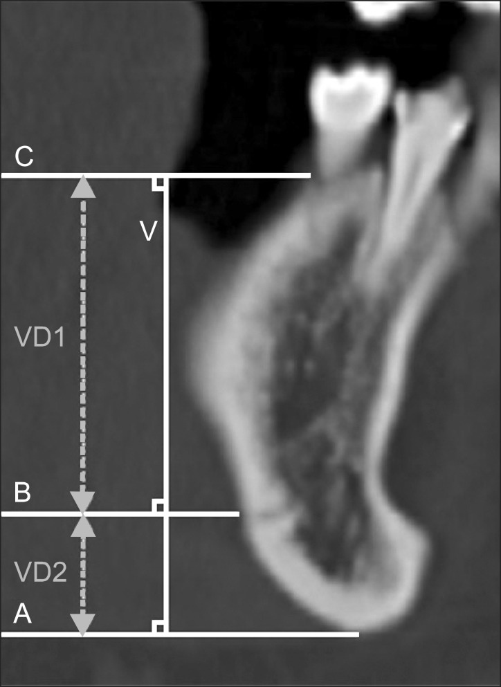
Fig. 1 Reference lines used in measurement on the coronal views of multi detector computed tomography. (C: third horizontal reference line, V: vertical reference line, B: second horizontal reference line, A: first horizontal reference line, VD1: vertical distance 1, VD2: vertical distance 2)
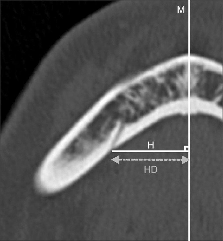
Fig. 2 Reference lines used in measurement on the axial views of multi detector computed tomography. (M: midline of mandible, H: vertical base line, HD: horizontal distance from midline to lingual foramina)
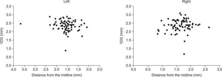
Fig. 3 Vertical distance from mandible border to lingual foramina (vertical distance 2, VD2) according to the distance from the midline.
Reference
-
1. Abrahams JJ, Hayt MW. Dental CT in pathologic changes of the maxillo-mandibular region. Radiologe. 1999; 39:1035–1043. PMID: 10643027.2. Dula K, Buser D, Porcellini B, Berthold H, Schwarz M. Computed tomography/oral implantology (I). Dental CT: a program for the computed tomographic imaging of the jaws: the principles and exposure technic. Schweiz Monatsschr Zahnmed. 1994; 104:450–459. PMID: 8209215.3. Angelopoulos C, Scarfe WC, Farman AG. A comparison of maxillofacial CBCT and medical CT. Atlas Oral Maxillofac Surg Clin North Am. 2012; 20:1–17. PMID: 22365427.
Article4. Laboda G. Life-threatening hemorrhage after placement of an endosseous implant: report of case. J Am Dent Assoc. 1990; 121:599–600. PMID: 2229738.
Article5. Mason ME, Triplett RG, Alfonso WF. Life-threatening hemorrhage from placement of a dental implant. J Oral Maxillofac Surg. 1990; 48:201–204. PMID: 2299461.
Article6. Mordenfeld A, Andersson L, Bergström B. Hemorrhage in the floor of the mouth during implant placement in the edentulous mandible: a case report. Int J Oral Maxillofac Implants. 1997; 12:558–561. PMID: 9274086.7. Kattan B, Snyder HS. Lingual artery hematoma resulting in upper airway obstruction. J Emerg Med. 1991; 9:421–424. PMID: 1787287.8. Kalpidis CD, Setayesh RM. Hemorrhaging associated with endosseous implant placement in the anterior mandible: a review of the literature. J Periodontol. 2004; 75:631–645. PMID: 15212344.
Article9. McDonnell D, Reza Nouri M, Todd ME. The mandibular lingual foramen: a consistent arterial foramen in the middle of the mandible. J Anat. 1994; 184:363–369. PMID: 8014127.10. Tepper G, Hofschneider UB, Gahleitner A, Ulm C. Computed tomographic diagnosis and localization of bone canals in the mandibular interforaminal region for prevention of bleeding complications during implant surgery. Int J Oral Maxillofac Implants. 2001; 16:68–72. PMID: 11280364.11. Gahleitner A, Hofschneider U, Tepper G, Pretterklieber M, Schick S, Zauza K, et al. Lingual vascular canals of the mandible: evaluation with dental CT. Radiology. 2001; 220:186–189. PMID: 11425994.
Article12. Yoshida S, Kawai T, Okutsu K, Yosue T, Takamori H, Sunohara M, et al. The appearance of foramen in the internal aspect of the mental region of mandible from Japanese cadavers and dry skulls under macroscopic observation and three-dimensional CT images. Okajimas Folia Anat Jpn. 2005; 82:83–87. PMID: 16350420.
Article13. Kawai T, Sato I, Yosue T, Takamori H, Sunohara M. Anastomosis between the inferior alveolar artery branches and submental artery in human mandible. Surg Radiol Anat. 2006; 28:308–310. PMID: 16547603.
Article14. Lustig JP, London D, Dor BL, Yanko R. Ultrasound identification and quantitative measurement of blood supply to the anterior part of the mandible. Oral Surg Oral Med Oral Pathol Oral Radiol Endod. 2003; 96:625–629. PMID: 14600700.
Article15. Liang X, Jacobs R, Lambrichts I, Vandewalle G, van Oostveldt D, Schepers E, et al. Microanatomical and histological assessment of the content of superior genial spinal foramen and its bony canal. Dentomaxillofac Radiol. 2005; 34:362–368. PMID: 16227480.
Article16. Tagaya A, Matsuda Y, Nakajima K, Seki K, Okano T. Assessment of the blood supply to the lingual surface of the mandible for reduction of bleeding during implant surgery. Clin Oral Implants Res. 2009; 20:351–355. PMID: 19298289.
Article17. Shiller WR, Wiswell OB. Lingual foramina of the mandible. Anat Rec. 1954; 119:387–390. PMID: 13197797.
Article18. Kim HJ, Choi BY, Lee HY, Chung IH. Morphological study of the mental spine, lingual foramen and nutrient foramen and innominate foramen in Korean mandibles. Korean J Phys Anthropol. 1993; 6:129–140.
Article19. Loukas M, Kinsella CR Jr, Kapos T, Tubbs RS, Ramachandra S. Anatomical variation in arterial supply of the mandible with special regard to implant placement. Int J Oral Maxillofac Surg. 2008; 37:367–371. PMID: 18262766.
Article20. Liang X, Jacobs R, Lambrichts I, Vandewalle G. Lingual foramina on the mandibular midline revisited: a macroanatomical study. Clin Anat. 2007; 20:246–251. PMID: 16683247.
Article21. Krenkel C, Holzner K. Lingual bone perforation as causal factor in a threatening hemorrhage of the mouth floor due to a single tooth implant in the canine region. Quintessenz. 1986; 37:1003–1008. PMID: 3489246.22. Isaacson TJ. Sublingual hematoma formation during immediate placement of mandibular endosseous implants. J Am Dent Assoc. 2004; 135:168–172. PMID: 15005432.
Article23. Budihardja AS, Pytlik C, Haarmann S, Holzle F. Hemorrhage in the floor of the mouth after second-stage surgery: case report. Implant Dent. 2006; 15:148–152. PMID: 16766897.
Article24. Niamtu J 3rd. Near-fatal airway obstruction after routine implant placement. Oral Surg Oral Med Oral Pathol Oral Radiol Endod. 2001; 92:597–600. PMID: 11740473.
Article25. Ferneini E, Gady J, Lieblich SE. Floor of mouth hematoma after posterior mandibular implants placement: a casereport. J Oral Maxillofac Surg. 2009; 67:1552–1554. PMID: 19531435.26. Givol N, Chaushu G, Halamish-Shani T, Taicher S. Emergency tracheostomy following life-threatening hemorrhage in the floor of the mouth during immediate implant placement in the mandibular canine region. J Periodontol. 2000; 71:1893–1895. PMID: 11156047.
Article27. Mardinger O, Manor Y, Mijiritsky E, Hirshberg A. Lingual perimandibular vessels associated with life-threatening bleeding: an anatomic study. Int J Oral Maxillofac Implants. 2007; 22:127–131. PMID: 17340906.28. Kalpidis CD, Konstantinidis AB. Critical hemorrhage in the floor of the mouth during implant placement in the first mandibular premolar position: a case report. Implant Dent. 2005; 14:117–124. PMID: 15968182.
Article29. Woo BM, Al-Bustani S, Ueeck BA. Floor of mouth haemorrhage and life-threatening airway obstruction during immediate implant placement inthe anterior mandible. Int J Oral Maxillofac Surg. 2006; 35:961–964. PMID: 16829038.30. Goldstein BH. Acute dissecting hematoma: a complication of oral and maxillofacial surgery. J Oral Surg. 1981; 39:40–43. PMID: 6969794.31. Dreiseidler T, Mischkowski RA, Neugebauer J, Ritter L, Zöller JE. Comparison of cone-beam imaging with orthopantomography and computerized tomography for assessment in presurgical implant dentistry. Int J Oral Maxillofac Implants. 2009; 24:216–225. PMID: 19492636.
- Full Text Links
-
- Actions
-
Cited
- CITED
-
- Close
- Share
-
- Similar articles
-
- Anatomical structure of lingual foramen in cone beam computed tomography
- Evaluation of mandibular cortical bone thickness for placement of temporary anchorage devices (TADs)
- Mandibular anatomy related to sagittal split ramus osteotomy in Koreans
- CBCT study of mandibular first molars with a distolingual root in Koreans
- A computerized tomographic study on the location of the mandibular canal and the cortical thickness of the mandible
