Comparison of the Pollard Method and the MRI Dimensions for Meniscal Sizing in Koreans
- Affiliations
-
- 1Department of Orthopaedic Surgery, Youngdong Severance Hospital, Yonsei University College of Medicine, Seoul, Korea. SWOOSUK@yuhs.ac
- KMID: 1947780
- DOI: http://doi.org/10.4055/jkoa.2007.42.6.743
Abstract
-
PURPOSE: The purpose of this study was to investigate the accuracy of the Pollard method for meniscal sizing of the meniscal allograft by comparison with the MRI dimensions.
MATERIALS AND METHODS
The width and length of 50 medial and lateral menisci were measured and compared using the Pollard method and MRI. The meniscal thickness was measured using MRI and we evaluated the individual differences.
RESULTS
The measurements of the width of the medial meniscus and the length of the lateral meniscus using the Pollard method and MRI were similar (p=0.459, p=0.108, respectively). However, the measurements of the length of the medial meniscus and the width of the lateral meniscus using MRI were significantly higher that those measured using the Pollard method (p=0.000 and p=0.001, respectively). The medial and lateral meniscal thicknesses were 6.26+/-0.86 mm and 6.47+/-0.84 mm, respectively, and there was no significant individual difference.
CONCLUSION
The measurements of the length of the medial meniscus and the width of the lateral meniscus, using the Pollard method and on MRI had significant differences. The Pollard method must be modified for meniscal sizing.
Keyword
Figure
-
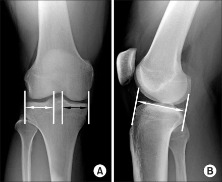
Fig. 1 The Pollard method. (A) An anteroposterial radiograph of the knee. The medial meniscal width is measured from the peak of the medial tibial eminence to the medial tibial metaphyseal margin(black arrow) and the lateral meniscal width is measured from the peak of the lateral tibial eminence to the periphery of the lateral tibial metaphysic(white arrow). (B) An lateral radiograph of the knee. The medial meniscal length is 80% of the sagittal tibial plateau distance (white arrow) measured at the joint line between a line parallel to the anterior tibia above the tuberosity and one tangent to the posterior plateau margin perpendicular to the joint line. The lateral meniscal length is 70% of the sagittal tibial plateau distance.

Fig. 2 MRI meniscal dimensions. (A) The medial meniscal width is measured from the widest medial meniscal width of the MRI coronal view. (B) The medial meniscal length is measured from the longest medial meniscal length of the MRI sagittal view. (C) The lateral meniscal width is measured from the widest lateral meniscal width of the MRI coronal view. (D) The lateral meniscal length is measured from the longest lateral meniscal length of the MRI sagittal view. (E) The meniscal height is measured from the highest height of the MRI sagittal view.
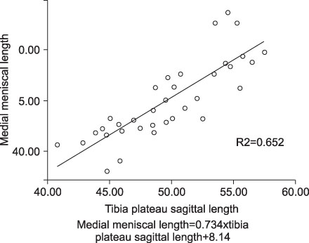
Fig. 3 The graph of simple linear regression analysis between the tibia plateau sagittal length and the medial meniscal length.
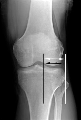
Fig. 4 An anteroposterial radiograph of the knee. The periphery of the lateral tibial metaphysis is round shaped, so there is a significant difference between the lateral epiphyseal margin and the lateral metaphyseal margin.
Reference
-
1. Carpenter JE, Wojtys EM, Huston LJ, Crabbe JP, Aisen AM. Preoperative sizing of meniscal allografts. Arthroscopy. 1993. 9:344.2. Dienst M, Greis PE, Ellis BJ, Bachus KN, Burks RT. Effect of lateral meniscal allograft sizing on contact mechanics of the lateral tibial plateau: an experimental study in human cadaveric knee joints. Am J Sports Med. 2007. 35:34–42.3. Fairbank TJ. Knee joint changes after meniscectomy. J Bone Joint Surg Br. 1948. 30:664–670.
Article4. Haut TL, Hull ML, Howell SM. Use of roentgenography and magnetic resonance imaging to predict meniscal geometry determined with a three-dimensional coordinate digitizing system. J Orthop Res. 2000. 18:228–237.
Article5. Higuchi H, Kimura M, Shirakura K, Terauchi M, Takagishi K. Factors affecting long-term results after arthroscopic partial meniscectomy. Clin Orthop Relat Res. 2000. 377:161–168.
Article6. Hoser C, Fink C, Brown C, Reichkendler M, Hackl W, Bartlett J. Long-term results of arthroscopic partial lateral meniscectomy in knees without associated damage. J Bone Joint Surg Br. 2001. 83:513–516.
Article7. Johnson DL, Swenson TM, Livesay GA, Aizawa H, Fu FH, Harner CD. Insertion-site anatomy of the human menisci: gross, arthroscopic, and topographical anatomy as a basis for meniscal transplantation. Arthroscopy. 1995. 11:386–394.
Article8. Johnson RJ, Kettelkamp DB, Clark W, Leaverton P. Factors effecting late results after meniscectomy. J Bone Joint Surg Am. 1974. 56:719–729.9. Kohn D, Moreno B. Meniscus insertion anatomy as a basis for meniscus replacement: a morphological cadaveric study. Arthroscopy. 1995. 11:96–103.
Article10. Lazović D, Wirth CJ, Knösel T, Gossé F, Maschek HG. Meniscus replacement using incongruent transplants--an experimental study. Z Orthop Ihre Grenzgeb. 1997. 135:131–137.11. Lubowitz JH, Verdonk PC, Reid JB 3rd, Verdonk R. Meniscus allograft transplantation: a current concepts review. Knee Surg Sports Traumatol Arthrosc. 2007. 15:476–492.
Article12. Matava MJ. Meniscal allograft transplantation: a systematic review. Clin Orthop Relat Res. 2007. 455:142–157.13. McDermott ID, Sharifi F, Bull AM, Gupte CM, Thomas RW, Amis AA. An anatomical study of meniscal allograft sizing. Knee Surg Sports Traumatol Arthrosc. 2004. 12:130–135.
Article14. Pollard ME, Kang Q, Berg EE. Radiographic sizing for meniscal transplantation. Arthroscopy. 1995. 11:684–687.
Article15. Rodeo SA. Meniscal allografts--where do we stand? Am J Sports Med. 2001. 29:246–261.
Article16. Sekaran SV, Hull ML, Howell SM. Nonanatomic location of the posterior horn of a medial meniscal autograft implanted in a cadaveric knee adversely affects the pressure distribution on the tibial plateau. Am J Sports Med. 2002. 30:74–82.
Article17. Shaffer B, Kennedy S, Klimkiewicz J, Yao L. Preoperative sizing of meniscal allografts in meniscus transplantation. Am J Sports Med. 2000. 28:524–533.
Article18. Stone KR, Stoller DW, Irving SG, Elmquist C, Gildengorin G. 3D MRI volume sizing of knee meniscus cartilage. Arthroscopy. 1994. 10:641–644.
Article19. Tapper EM, Hoover NW. Late results after meniscectomy. J Bone Joint Surg Am. 1969. 51:517–526.
Article20. Veltri DM, Warren RF, Wickiewicz TL, O'Brien SJ. Current status of allograft meniscal transplantation. Clin Orthop Relat Res. 1994. 44–55.
Article21. Verstraete KL, Verdonk R, Lootens T, Verstraete P, De Rooy J, Kunnen M. Current status and imaging of allograft meniscal transplantation. Eur J Radiol. 1997. 26:16–22.
Article22. Wilcox TR, Goble EM. Indications for meniscal allograft reconstruction. Am J Knee Surg. 1996. 9:35–36.23. Wilcox TR, Goble EM, Doucette SA. Goble technique of meniscus transplantation. Am J Knee Surg. 1996. 9:37–42.24. Yocum LA, Kerlan RK, Jobe FW, et al. Isolated lateral meniscectomy. A study of twenty-six patients with isolated tears. J Bone Joint Surg Am. 1979. 61:338–342.
Article
- Full Text Links
-
- Actions
-
Cited
- CITED
-
- Close
- Share
-
- Similar articles
-
- Operative Verification of Meniscal Injury in Magnetic Resonance Imaging
- Ananalysis of the Clinical and MRI Findings of the Bucket: Handle Meniscal Tears of the Knee Joint
- MR Findings of Meniscal Cysts
- MR imaging in meniscus injury of knee joint
- A comparison of Bone SPECT, MRI, and Arthroscopy for the Evaluation of Chronic Knee Pain
