J Menopausal Med.
2013 Dec;19(3):139-142. 10.6118/jmm.2013.19.3.139.
A Case of Vaginal Cancer with Uterine Prolapse
- Affiliations
-
- 1Department of Obstetrics and Gynecology, Pusan National University School of Medicine, Yangsan, Korea. ohchoi@pusan.ac.kr
- KMID: 1939538
- DOI: http://doi.org/10.6118/jmm.2013.19.3.139
Abstract
- Primary vaginal cancer combined with uterine prolapse is very rare. We present a case of 80-year-old postmenopausal women complaints of something coming out per vagina for the past 20 years, along with blood stained discharge, foul odor leukorrhea, and severe pelvic pain for the last 3 months. A 4 x 5 cm ulcer was present on middle third of vaginal wall with marked edema and ulceration of surrounding tissue. The prolapse was reduced under intravenous sedation in operating room. On gynecologic examination, uterus was normal in size, no adnexal mass was examined, and both parametrium were thickened. Papanicolaou smear was normal. Biopsy of the ulcer at vaginal wall revealed invasive squamous cell carcinoma of vagina. Magnetic Resonance Imaging of abdomen and pelvis showed left hydronephrosis and liver metastasis. Positron emission tomography (PET)/computed tomography (CT) revealed metastasis to lung, liver and iliac bone. She died from progression of disease one month after diagnosis.
Keyword
MeSH Terms
-
Abdomen
Aged, 80 and over
Biopsy
Blood Stains
Carcinoma, Squamous Cell
Diagnosis
Edema
Female
Humans
Hydronephrosis
Leukorrhea
Liver
Lung
Magnetic Resonance Imaging
Neoplasm Metastasis
Odors
Operating Rooms
Papanicolaou Test
Pelvic Pain
Pelvis
Positron-Emission Tomography
Postmenopause
Prolapse
Ulcer
Uterine Prolapse*
Uterus
Vagina
Vaginal Neoplasms*
Figure
-
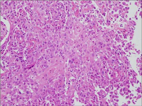
Fig. 1 Histopathology of biopsied specimen showing squamous cell carcinoma of the vagina (H & E, × 200).
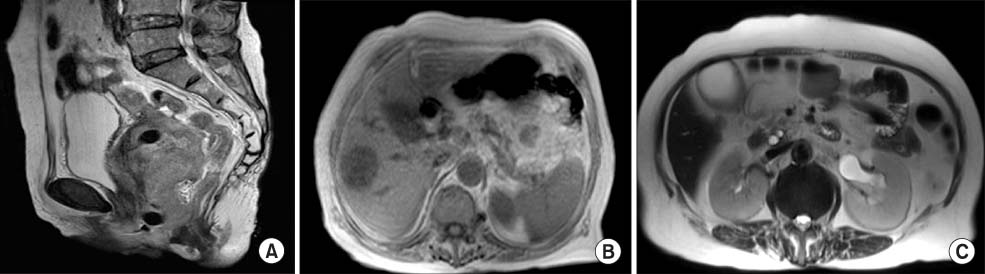
Fig. 2 (A) A high signal intensity lesion at posterior vagina in T2-weighted sagittal magnetic resonance imaging (MRI) image. (B) A liver metastasis in T2-weighted axial MRI image. (C) A Left hydronephrosis T2-weighted axial MRI image.
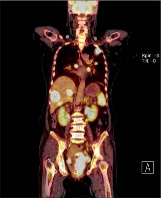
Fig. 3 Multiple hypermetabolic lesion at lung, liver and iliac bone in positron emission tomography/computed tomography.
Reference
-
1. Benedet JL, Bender H, Jones H 3rd, Ngan HY, Pecorelli S. FIGO Committee on Gynecologic Oncology. FIGO staging classifications and clinical practice guidelines in the management of gynecologic cancers. Int J Gynaecol Obstet. 2000; 70:209–262.2. Iavazzo C, Vorgias G, Vecchini G, Katsoulis M, Akrivos T. Vaginal carcinoma in a completely prolapsed uterus. A case report. Arch Gynecol Obstet. 2007; 275:503–505.3. Karateke A, Tugrul S, Yakut Y, Gürbüz A, Cam C. Management of a case of primary vaginal cancer with irreducible massive uterine prolapse--a case report. Eur J Gynaecol Oncol. 2006; 27:528–530.4. Creasman WT. Vaginal cancers. Curr Opin Obstet Gynecol. 2005; 17:71–76.5. Park J, Kim TH, Lee HH, Lee W, Chung SH. Lichen sclerosus in a post-menopausal woman: a case report. J Korean Soc Menopause. 2012; 18:70–73.6. Berek JS, Hacker NF. Berek and Hacker's gynecologic oncology. 5th ed. Philadelphia, PA: Lippincott Williams & Wilkins;2010.7. Barakat RR, Markman M, Randall ME. Principles and practice of gynecologic oncology. 5th ed. Philadelphia, PA: Lippincott Williams & Wilkins;2009.8. Howat JM, Stassan L, Mohandas I, Daw E. Carcinoma of the vagina presenting as a ruptured procidentia with an entero-vaginal fistula and prolapse of the small bowel. Postgrad Med J. 1984; 60:435–436.9. Ghosh SB, Tripathi R, Mala YM, Khurana N. Primary invasive carcinoma of vagina with third degree uterovaginal prolapse: a case report and review of literature. Arch Gynecol Obstet. 2009; 279:91–93.10. Gupta N, Mittal S, Dalmia S, Misra R. A rare case of primary invasive carcinoma of vagina associated with irreducible third degree uterovaginal prolapse. Arch Gynecol Obstet. 2007; 276:563–564.11. Rao K, Kumar NP, Geetha AS. Primary carcinoma of vagina with uterine prolapse. J Indian Med Assoc. 1989; 87:10–12.12. Fonseca AM, Pereyra EAG, Valente SE, Assoni L, Souza AZ. Colposcopic, cytologic and histologic findings in patients with uterine prolapse of second and third degree. Arq Bras Med. 1988; 62:273–275.13. Cho KI, Prk CH, Choi GS, Huh CK. To cases of uterine prolapse combined with cervical carcinoma. Korean J Obstet Gynecol. 1993; 36:3351–3357.
- Full Text Links
-
- Actions
-
Cited
- CITED
-
- Close
- Share
-
- Similar articles
-
- Efficacy of Posterior IVS for the Patients with Vaginal Vault and Uterine Prolapse
- A Case Report of Rectal Herniation through Rectovaginal Fistula Associated with Uterine Prolapse
- A Case of Uterine Prolapse in Pregnancy
- A Case Report of Cervical Prolapse Complicating Pregnancy
- Clinical study of the sacrospinous ligament suspension using Miya hook in management of pelvic organ prolapse
