Prog Med Phys.
2013 Sep;24(3):183-190. 10.14316/pmp.2013.24.3.183.
Development of Photoacoustic System for Breast Cancer Detection
- Affiliations
-
- 1Department of Medical Science, School of Medicine, Ewha Womans University, Seoul, Korea.
- 2Department of Radiation Oncology, School of Medicine, Ewha Womans University, Seoul, Korea. renalee@ewha.ac.kr
- KMID: 1910583
- DOI: http://doi.org/10.14316/pmp.2013.24.3.183
Abstract
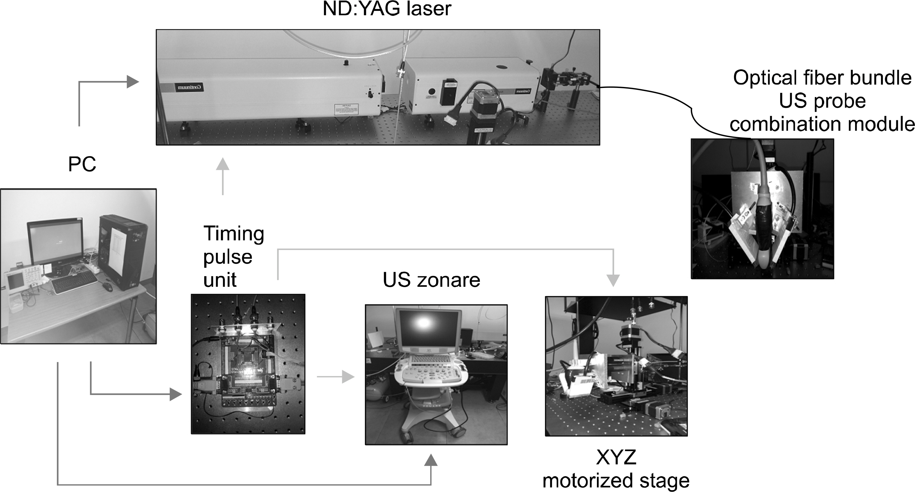
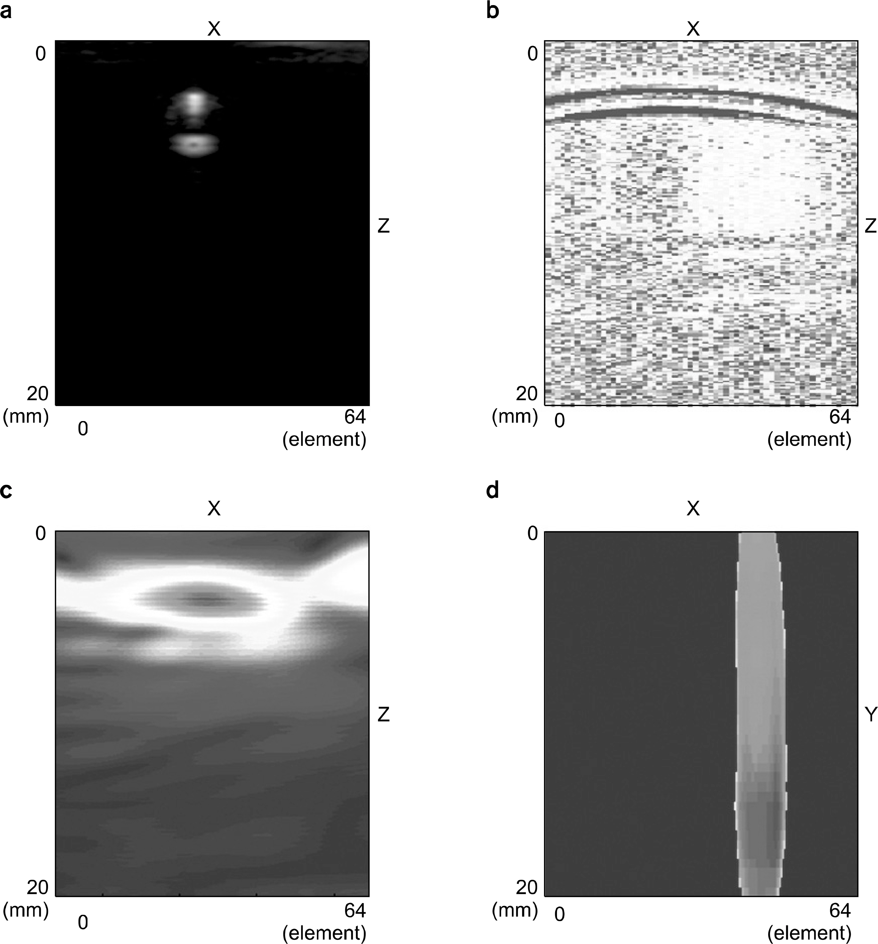
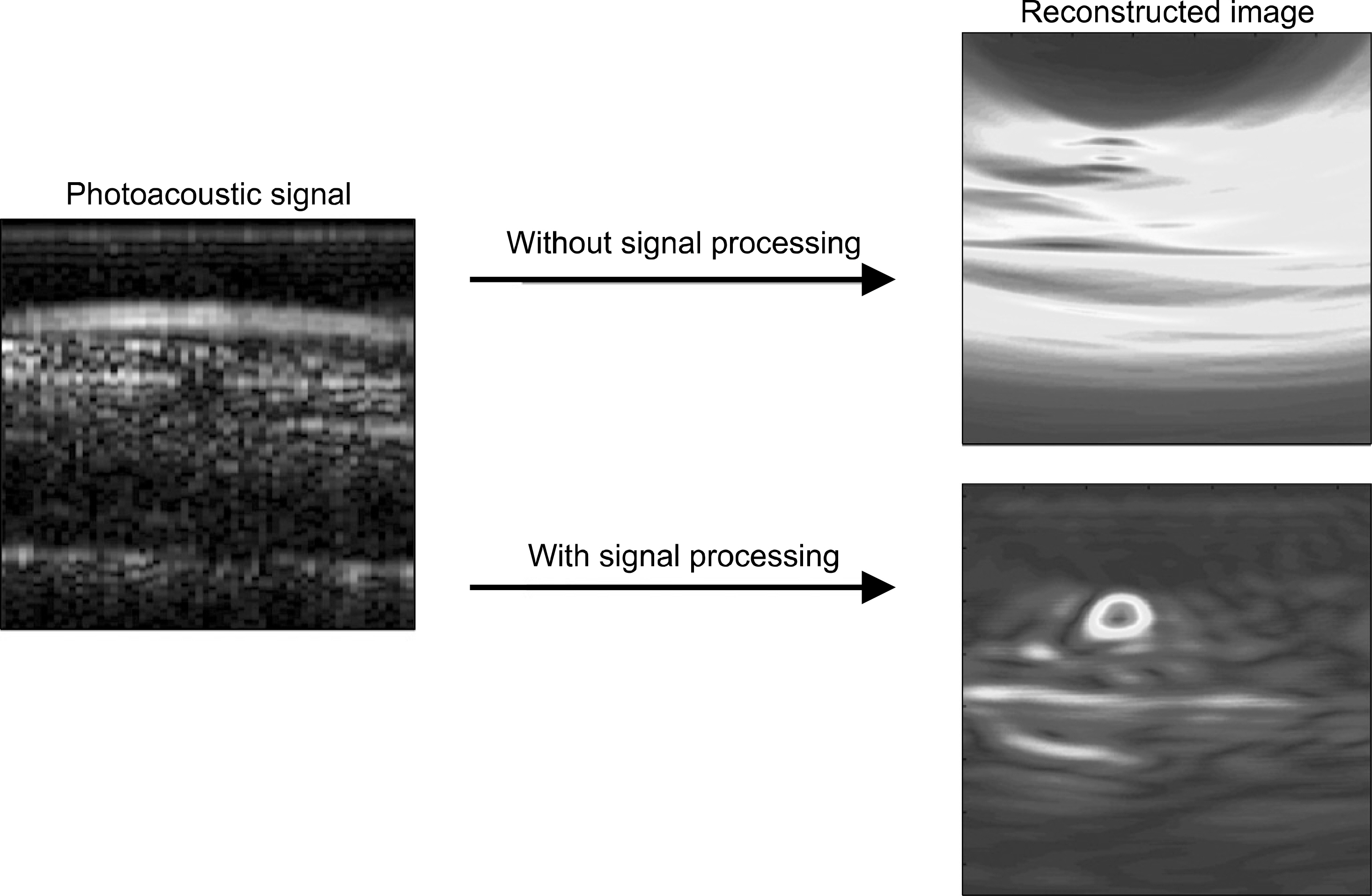
- Recently, the photoacoustic imaging system has been widely and intensively developed, and has been shown the possibility of diagnosis for early stage cancer. In this study, we developed a photoacoustic tomography imaging system with a commercial ultra sound device and a linear array probe. A tube phantom and a chicken breast phantom was made for the possibility of a system as a breast cancer detection. A moving average filter and a band pass filter with 3~6 MHz bandwidth were developed for background noise elimination before delay-and-sum beamforming algorithm was used for image reconstruction. As a result, we showed that some signal processing procedure before beamforming was effective for the photoacoustic image reconstruction.
Figure
Reference
-
1. Xu M, Wang LH. Photoacoustic imaging in biomedicine. Review of Scientific Instruments. 77(4):041101. 2006.
Article2. Li C, Wang LH. Photoacoustic tomography and sensing in biomedicine Phys Med Biol. 54(19):59–97. 2009.3. Weidner N, Semple JP, Welch WR, Folkman J. Tumor angiogenesis and metastasis–correlation in invasive breast carcinoma. N Engl J Med. 324(1):1–8. 1991.4. Heijblom M, Piras D, Xia W, et al. Visualizing breast cancer using the Twente photoacoustic mammoscope: What do we learn from twelve new patient measurements? Opt Express. 20(11):11582–11597. 2012.
Article5. 김주혜, 허장용, 오정환 등. 유방암 진단용 광음향 영상 시스템 특성 평가를 위항 팬텀 개발. 의학물리. 23(1):28–30. 2012.6. Park SH, Aglyamov SR, Emelianov SY. Beamforming for photoacoustic imaging using linear array transducer. IEEE Ultrasonics Symposium 856-859. 2007.7. Kruger RA, Kiser WL, Reinecke DR, Kruger GA. Thermoacoustic computed tomography using a conventional linear transducer array. Medical Physics. 30(5):856–860. 2003.
Article8. Ku G, Wang X, Stoica G, Wang LH. Multiple-bandwidth photoacoustic tomography. Phys Med Biol. 49(7):1329–38. 2004.
Article9. Duck FA. Physical Properties of Tissue. Academic Press;London: 1990. ), pp.p. 120–130.10. Wells PNT. Ultrasonic imaging of the human body. Rep Prog Phys. 62(5):671. 1999.
Article
- Full Text Links
- Actions
-
Cited
- CITED
-
- Close
- Share
- Similar articles
-
- Development and clinical translation of photoacoustic mammography
- Clinical photoacoustic imaging of cancer
- Review of Photoacoustic Imaging for Imaging-Guided Spinal Surgery
- Photoacoustic imaging of occlusal incipient caries in the visible and near-infrared range
- Fast photoacoustic imaging systems using pulsed laser diodes: a review


