Chonnam Med J.
2014 Apr;50(1):21-22. 10.4068/cmj.2014.50.1.21.
Medullary Sponge Kidney on Retrograde Pyelography
- Affiliations
-
- 1Department of Urology, Kaohsiung Medical University Hospital, Kaohsiung Medical University, Kaohsiung, Taiwan. shpihu@yahoo.com.tw
- 2School of Post-baccalaureate Medicine, Kaohsiung Medical University, Kaohsiung, Taiwan.
- 3Department of Urology, Faculty of Medicine, College of Medicine, Kaohsiung Medical University, Kaohsiung, Taiwan.
- KMID: 1889758
- DOI: http://doi.org/10.4068/cmj.2014.50.1.21
Abstract
- A woman aged 31 had recurrent urinary tract infection with bloody urine. A series image of medullary sponge kidney presented by intravenous urography (IVU) was detected dynamically by retrograde pyelography (RP). Other than ultrasonography and IVU, RP is also a reliable method to detect medullary sponge kidney.
Figure
-
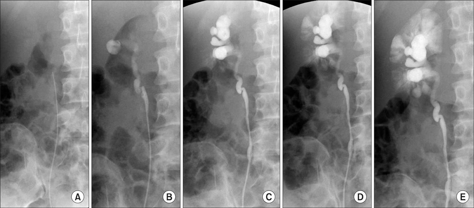
FIG. 1 Retrograde pyelography (RP).
Reference
-
1. Katabathina VS, Kota G, Dasyam AK, Shanbhogue AK, Prasad SR. Adult renal cystic disease: a genetic, biological, and developmental primer. Radiographics. 2010; 30:1509–1523.
Article2. Hida T, Nishie A, Asayama Y, Ishigami K, Fujita N, Inokuchi J, et al. MR imaging of focal medullary sponge kidney: case report. Magn Reson Med Sci. 2012; 11:65–69.
Article
