An Unusual Variant of Anlage Tumor of Pineal Region in an Infant
- Affiliations
-
- 1Department of Neurosurgery, King Edward Memorial Hospital and Seth Gordhandas Sunderdas Medical College, Mumbai, India. drraghavr@gmail.com
- 2Department of Neuropathology, King Edward Memorial Hospital and Seth Gordhandas Sunderdas Medical College, Mumbai, India.
- KMID: 1882012
- DOI: http://doi.org/10.14791/btrt.2015.3.1.52
Abstract
- A 9-month-old male child was brought with complaints of increasing head size for 2 months, increasing lethargy and vomiting for the last 2 days. Radiology revealed a heterogeneously enhancing, globular lesion in the pineal region with hydrocephalus. Near total excision of the tumor was carried out. The histopathological examination of the lesion showed heterogenous elements in the form of mature neuroepithelial and ectomesenchymal tissue. The pathology and radiology of this unusual lesion is discussed with relevant review of literature.
Keyword
MeSH Terms
Figure
-
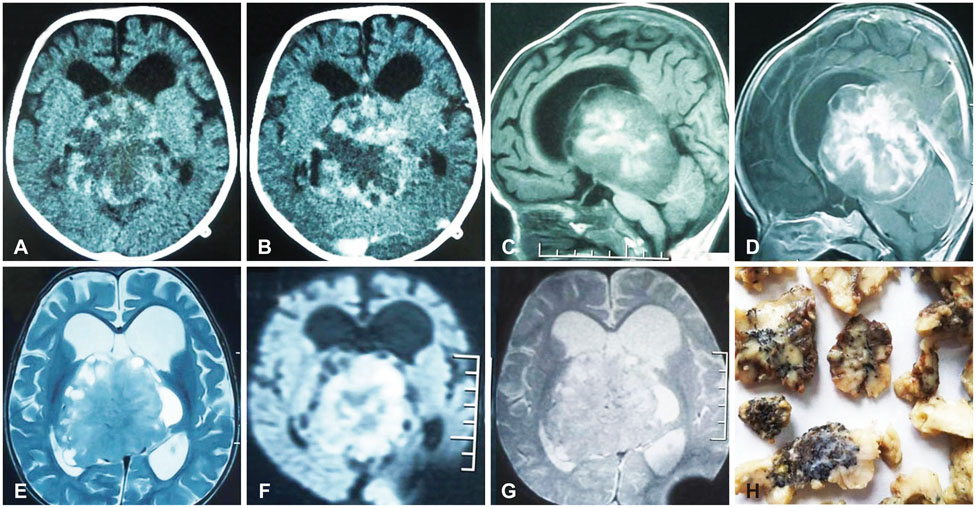
Fig. 1 Radiology of the tumor. A: Plain computed tomography of brain showing a well-defined, heterogenous lesion in the pineal region. B: Post contrast computed tomography showing patchy heterogenous enhancement of the lesion. Magnetic resonance (MR) imaging showed mixed intensity on T1 (C), T2 (D) weighted sequences with peripheral cystic areas and heterogeneous enhancement (E). Diffusion weighted MR image revealed restricted diffusion suggestive of high cellularity (F). Gradient recovery echo sequence of MR showing absence of blooming (G). Specimen of tumor showing multiple firm, white glistening pieces with punctate brown black pigmentation on cut surface suggestive of melanin (H).
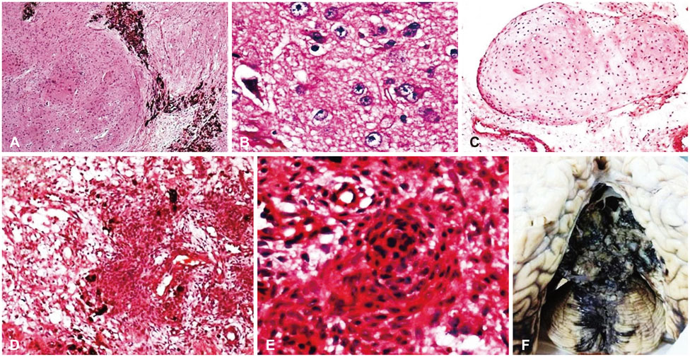
Fig. 2 Histopathology of the tumor. A: Nodules of neuroglial tissue separated by septa with nested pigmented neuroepithelial tubules (H&E, ×40). B: Glial cells and ganglion cells, one binucleate ganglion cell is seen (H&E, ×400). C: Mature hyaline cartilage (H&E, ×400). D: Meningioangiomatosis areas (H&E, ×100). E: Whorling pattern of spindle cells suggestive of meningiomatosis (H&E, ×400). F: Autopsy specimen of brain showing shell of residual tumor anterolaterally.
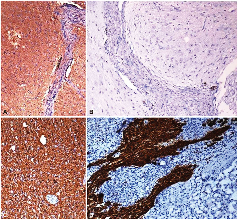
Fig. 3 Immunohistochemistry of the tumor. A: Synaptophysin positivity (×100). B: Mib1 labelling index <1% (×100). C and D: Glial fibrillary acidic protein positive in the neuroglial nodules and negative in the meningioangiomatosis areas (×100).
Reference
-
1. Ajayi O, Palma A, Sadanand V, Deisch J. Pineal anlage tumor: case report and review of literature. JSM Neurosurg Spine. 2014; 2:1035.2. Fuller GN, Scheithauer BW. The 2007 Revised World Health Organization (WHO) Classification of Tumours of the Central Nervous System: newly codified entities. Brain Pathol. 2007; 17:304–307.
Article3. Gudinaviciene I, Pranys D, Zheng P, Kros JM. A 10-month-old boy with a large pineal tumor. Brain Pathol. 2005; 15:263–264.
Article4. Yi JW, Kim HJ, Choi YJ, et al. Successful treatment by chemotherapy of pineal parenchymal tumor with intermediate differentiation: a case report. Cancer Res Treat. 2013; 45:244–249.
Article5. Schmidbauer M, Budka H, Pilz P. Neuroepithelial and ectomesenchymal differentiation in a primitive pineal tumor ("pineal anlage tumor"). Clin Neuropathol. 1989; 8:7–10.6. Berns S, Pearl G. Review of pineal anlage tumor with divergent histology. Arch Pathol Lab Med. 2006; 130:1233–1235.
Article
