Multiple variations in the branches of the coeliac trunk
- Affiliations
-
- 1Department of Anatomy, Kasturba Medical College, Manipal University, Manipal, India. sushma.rk@manipal.edu
- 2Department of Anatomy, Father Muller Medical College, Mangalore, India.
- KMID: 1845284
- DOI: http://doi.org/10.5115/acb.2015.48.2.147
Abstract
- Here we present a unique case of variation in the branching pattern of the coeliac trunk. In the present case, the coeliac trunk was replaced by two separate arterial trunks. The first arterial trunk bifurcated into the left gastric and the left hepatic arteries. The second arterial trunk bifurcated into a splenic artery and a hepato-gastroduodenal trunk. The hepato-gastroduodenal trunk presented an unusual course and termination. The right hepatic artery arising from the hepato-gastroduodenal trunk also showed a variant course. Such rare variations are important for gastroenterological surgeons and interventional radiologists due to increase in number of transplantation surgeries and live donor liver transplantations.
Keyword
Figure
-
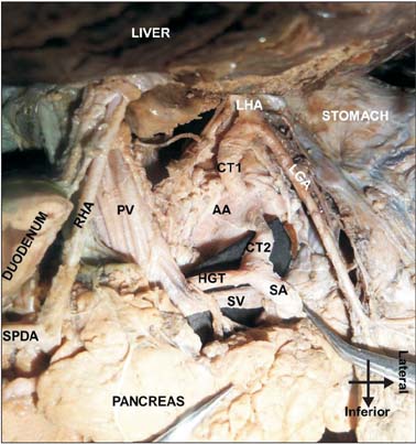
Fig. 1 Showing two separate arterial trunks (CT1 and CT2) at the level of lower border of T12 vertebra arising from the abdominal aorta (AA). A well-defined coeliac trunk was absent. The first arterial trunk (CT1), after arising from the abdominal aorta further bifurcated and provided the left gastric (LGA) and the left hepatic arteries (LHA). The second arterial trunk (CT2), about 1.5 cm long further bifurcated into a splenic artery (SA) and a hepato-gastroduodenal trunk (HGT). The HGT presented an unusual course as it moved to the right, further passing deep to the portal vein (PV). RHA, right hepatic artery; SPDA, superior pancreatico-duodenal artery; SV, splenic vein.

Fig. 2 Showing the hepato-gastroduodenal trunk (HGT) emerging anteriorly at the lower border of the neck of the pancreas. It then bifurcated into right hepatic (RHA) and superior pancreatico-duodenal arteries (SPDAs). The right hepatic artery further ascended, passing anterior to the neck of the pancreas to reach the porta-hepatis to supply the right lobe of the liver. The cystic branch (CA) was also provided by the right hepatic artery. The SPDA when traced passed anterior to the head of the pancreas to reach the pancreatico-duodenal groove. GB, gallbladder; SMA, superior mesentric artery; SMV, superior mesentric vein.
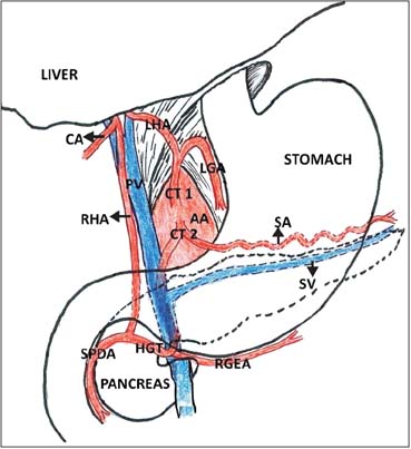
Fig. 3 Schematic representation of the variations in the branching pattern of the coeliac trunk. AA, abdominal aorta; CA, cystic artery; CT 1& 2, common trunks 1&2; HGT, hepatogastroduodenal trunk; LGA, left gastric artery; LHA, left hepatic artery; SA, splenic artery; SPDA, superior pancreatico-duodenal artery; SV, splenic vein; RGEA, right gastro-epiploeic artery; RHA, right hepatic artery; PV, portal vein.
Cited by 1 articles
-
A rare combined variation of the coeliac trunk, renal and testicular vasculature
Renate Elke Potgieter, Adam Michael Taylor, Quenton Wessels
Anat Cell Biol. 2018;51(1):62-65. doi: 10.5115/acb.2018.51.1.62.
Reference
-
1. Bergman RA, Afifi AK, Miyauchi R. Illustrated encyclopedia of human anatomic variation [Internet]. Anatomy Atlases;c1995-2015. cited 2015 Mar 1. Available from: http://www.anatomyatlases.org/AnatomicVariants/AnatomyHP.shtml.2. Vandamme JP, Bonte J. The branches of the celiac trunk. Acta Anat (Basel). 1985; 122:110–114.3. Saeed M, Murshid KR, Rufai AA, Elsayed SE, Sadiq MS. Coexistence of multiple anomalies in the celiac-mesenteric arterial system. Clin Anat. 2003; 16:30–36.4. Standring S, Borley NR, Collins P, Crossman AR, Gatzoulis MA, Healy JC, Johnson D, Mahadevan V, Newell RL, Wigley CB. Gray's anatomy: the anatomical basis of clinical practice. 40th ed. Edinburgh: Elsevier;2008. p. 1072–1074. p. 1379–1380.5. Yildirim M, Ozan H, Kutoglu T. Left gastric artery originating directly from the aorta. Surg Radiol Anat. 1998; 20:303–305.6. Saga T, Hirao T, Kitashima S, Watanabe K, Nohno M, Araki Y, Kobayashi S, Yamaki K. An anomalous case of the left gastric artery, the splenic artery and hepato-mesenteric trunk independently arising from the abdominal aorta. Kurume Med J. 2005; 52:49–52.7. Chaudhari ML, Maheria PB, Nerpagar S, Menezes VR. Origin of left accessory hepatic artery from the left gastric artery. Natl J Integr Res Med. 2013; 4:173–174.8. Daseler EH, Anson BJ. The cystic artery and constituents of the hepatic pedicle; a study of 500 specimens. Surg Gynecol Obstet. 1947; 85:47–63.9. Michels NA. Variational anatomy of the hepatic, cystic, and retroduodenal arteries; a statistical analysis of their origin, distribution, and relations to the biliary ducts in two hundred bodies. AMA Arch Surg. 1953; 66:20–34.10. Michels NA. Newer anatomy of the liver and its variant blood supply and collateral circulation. Am J Surg. 1966; 112:337–347.11. Hiatt JR, Gabbay J, Busuttil RW. Surgical anatomy of the hepatic arteries in 1000 cases. Ann Surg. 1994; 220:50–52.12. Ghosh SK. Variations in the origin of middle hepatic artery: a cadaveric study and implications for living donor liver transplantation. Anat Cell Biol. 2014; 47:188–195.13. Adachi B. Das Arteriensystem der Japaner. Band II. Kyoto: Verlag der Kaiserlich-Japanischen Universität zu Kyoto;1928.14. Ramanadham S, Toomay SM, Yopp AC, Balch GC, Sharma R, Schwarz RE, Mansour JC. Rare hepatic arterial anatomic variants in patients requiring pancreatoduodenectomy and review of the literature. Case Rep Surg. 2012; 2012:953195.15. Varotti G, Gondolesi GE, Munoz L, Florman S, Fishbein TM, Emre S, Schwartz ME, Miller C. 43: Biliary complications in 96 right lobe living donor liver transplants. J Gastrointest Surg. 2003; 7:271.
- Full Text Links
-
- Actions
-
Cited
- CITED
-
- Close
- Share
-
- Similar articles
-
- A rare combined variation of the coeliac trunk, renal and testicular vasculature
- Variations in the branching pattern of the celiac trunk and its clinical significance
- Study on branching pattern of aortic arch in Indian
- Double Facial Nerve Trunk Emerged from the Stylomastoid Foramen and Petrotympanic Fissure: A Case Report
- Anatomical Variation of the Glissonean Pedicle of the Right Liver
