Usefulness of Computed Tomography Hounsfield Unit Measurement for Diagnosis of Congenital Cholesteatoma
- Affiliations
-
- 1Department of Radiology, Medical Research Institute, Pusan National University Yangsan Hospital, College of Medicine, Pusan National University, Yangsan, Korea. kyw47914@gmail.com
- 2Department of Otolaryngology, Medical Research Institute, Pusan National University Yangsan Hospital, College of Medicine, Pusan National University, Yangsan, Korea.
- KMID: 1839425
- DOI: http://doi.org/10.3348/jksr.2014.70.2.153
Abstract
- PURPOSE
To evaluate the usefulness of Hounsfield unit (HU) measurements for diagnosing of congenital cholesteatoma.
MATERIALS AND METHODS
A total of 43 patients who underwent surgery due to middle ear cavity lesions were enrolled. Twenty-one patients were confirmed to have congenital cholesteatoma by histopathological results and the other 22 patients were confirmed to have otitis media (OM) by operation. Their computed tomography images were retrospectively reviewed. We measured HU of the soft tissue mass in the middle ear cavity. In addition, we evaluated the largest diameter and location of the mass, the presence of bony erosion in the ear ossicle, and the status of the tympanic membrane in the cholesteatoma group.
RESULTS
The mean HU was 37.36 +/- 6.11 (range, 27.5-52.5) in the congenital cholesteatoma group and 76.09 +/- 8.74 (range, 58.5-96) in the OM group (p < 0.001). The cut-off value was 55.5. The most common location for congenital cholesteatoma was the mesotympanum, and ear ossicle erosion was present in 24%. All patients had an intact tympanic membrane.
CONCLUSION
HU measurement may be useful as an additional indicator to diagnose congenital cholesteatoma.
MeSH Terms
Figure
-

Fig. 1 Congenital cholesteatoma of the left middle ear cavity in a 3-year-old boy. Axial (A) and reformatted coronal (B) temporal bone CT scans demonstrating a well-defined, round soft-tissue mass in the mesotympanum of the left middle ear cavity. The measured Hounsfield unit (HU) is 36 in axial image and 33 in coronal image. The lower HU of the two measured value were selected in this case. This mass considered as congenital cholesteatoma.
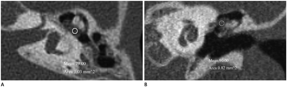
Fig. 2 Otitis media of the left middle ear cavity in a 5-year-old boy. Axial (A) and reformatted coronal (B) temporal bone CT scans demonstrating a well defined soft-tissue mass in epitympanum of the left middle ear cavity. It is difficult to distinguish otitis media from congenital cholesteatoma by image finding alone. The measured Hounsfield unit (HU) is 79 in axial image and 90 in coronal image. The lower HU of the two measured value were selected in this case. This mass considered as otitis media.

Fig. 3 The Hounsfield unit (HU) of congenital cholesteatoma group and otitis media group.
Reference
-
1. McDonald TJ, Cody DT, Ryan RE Jr. Congenital cholesteatoma of the ear. Ann Otol Rhinol Laryngol. 1984; 93(6 Pt 1):637–640.2. Trojanowska A, Trojanowski P, Olszanski W, Klatka J, Drop A. Differentiation between cholesteatoma and inflammatory process of the middle ear, based on contrast-enhanced computed tomography imaging. J Laryngol Otol. 2007; 121:444–448.3. De Foer B, Vercruysse JP, Bernaerts A, Meersschaert J, Kenis C, Pouillon M, et al. Middle ear cholesteatoma: non-echo-planar diffusion-weighted MR imaging versus delayed gadolinium-enhanced T1-weighted MR imaging--value in detection. Radiology. 2010; 255:866–872.4. Baráth K, Huber AM, Stämpfli P, Varga Z, Kollias S. Neuroradiology of cholesteatomas. AJNR Am J Neuroradiol. 2011; 32:221–229.5. Smith PG, Leonetti JP, Kletzker GR. Differential clinical and radiographic features of cholesterol granulomas and cholesteatomas of the petrous apex. Ann Otol Rhinol Laryngol. 1988; 97(6 Pt 1):599–604.6. Franklin DJ, Jenkins HA, Horowitz BL, Coker NJ. Management of petrous apex lesions. Arch Otolaryngol Head Neck Surg. 1989; 115:1121–1125.7. Park MH, Rah YC, Kim YH, Kim JH. Usefulness of computed tomography Hounsfield unit density in preoperative detection of cholesteatoma in mastoid ad antrum. Am J Otolaryngol. 2011; 32:194–197.8. Levenson MJ, Michaels L, Parisier SC. Congenital cholesteatomas of the middle ear in children: origin and management. Otolaryngol Clin North Am. 1989; 22:941–954.9. Phelps ME, Hoffman EJ, Ter-Pogossian MM. Attenuation coefficients of various body tissues, fluids, and lesions at photon energies of 18 to 136 keV. Radiology. 1975; 117(3 Pt 1):573–583.10. Brooks RA. A quantitative theory of the Hounsfield unit and its application to dual energy scanning. J Comput Assist Tomogr. 1977; 1:487–493.11. Caponetti G, Thompson LD, Pantanowitz L. Cholesteatoma. Ear Nose Throat J. 2009; 88:1196–1198.12. Fisch U. Tympanoplasty, Mastoidectomy, and Stapes Surgery. New York: Thieme;1994. p. 146.13. McGill TJ, Merchant S, Healy GB, Friedman EM. Congenital cholesteatoma of the middle ear in children: a clinical and histopathological report. Laryngoscope. 1991; 101(6 Pt 1):606–613.14. Nelson M, Roger G, Koltai PJ, Garabedian EN, Triglia JM, Roman S, et al. Congenital cholesteatoma: classification, management, and outcome. Arch Otolaryngol Head Neck Surg. 2002; 128:810–814.
- Full Text Links
-
- Actions
-
Cited
- CITED
-
- Close
- Share
-
- Similar articles
-
- Study for hounsfield units in computed tomogram with jaw lesion
- Congenital Cholesteatoma Localized to the Tip of the Mastoid Bone: A Case Report and Possible Etiology
- Congenital Cholesteatoma: Diagnosis and Management
- A Case of Simultaneous Presentation of Closed and Open Type of Congenital Cholesteatoma
- A Case of Two Isolated Congenital Cholesteatomas Presented in Middle Ear Cavity
