Prognostic significance of computed tomography defined ascites in advanced gastric cancer
- Affiliations
-
- 1Department of Surgery, Hallym University Kangdong Sacred Heart Hospital, Seoul, Korea. neosurgy@hallym.or.kr
- 2Department of Pathology, Hallym University Kangdong Sacred Heart Hospital, Seoul, Korea.
- 3Department of Radiology, Hallym University Kangdong Sacred Heart Hospital, Seoul, Korea.
- KMID: 1820069
- DOI: http://doi.org/10.4174/jkss.2012.82.4.219
Abstract
- PURPOSE
The aim of this study was to investigate the clinicopathologic features and prognosis in patients with computed tomography (CT) findings of ascites, with a focus on the correlation with peritoneal carcinomatosis.
METHODS
This study included a total of 157 patients who underwent surgery for advanced gastric cancer from 2003 to 2008 at the Department of Surgery, Hallym University Kangdong Sacred Heart Hospital, Seoul, Korea, which were analyzed retrospectively.
RESULTS
Fourteen patients (8.9%) presented ascites on their CT scan. Among them, 10 patients had peritoneal carcinomatosis, and showed significant difference with CT ascites positive group in the incidence of peritoneal carcinomatosis. The presence of CT ascites was significantly correlated with pathologic T stage, tumor size, histologic type, CT T and N stages, CT peritoneal nodularity and curability of surgery, statistically. The prognosis of CT ascites positive group was much poorer in the total advanced gastric cancer patients (P < 0.001), as well as in patients with pathologic T4 (P = 0.002). Also in patients without peritoneal carcinomatosis, CT ascites positive subgroup tended to have a worse prognosis than CT ascites negative subgroup (P = 0.086). Tumor size, CT T and N stages and the presence of CT peritoneal nodularity and ascites influenced the prognosis significantly; among which, if a tumor size larger than 5 cm, CT T4 stage and the presence of CT ascites were identified as independent prognostic factors.
CONCLUSION
The presence of ascites was closely associated with peritoneal metastasis, and was the most significant independent prognostic factor in advanced gastric cancer in the present study.
Keyword
MeSH Terms
Figure
-
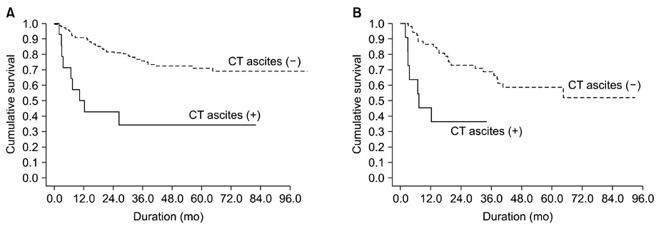
Fig. 1 (A) Cumulative overall survival of 157 advanced gastric cancer patients according to the status of computed tomography (CT) ascites (P < 0.001) (log rank test). (B) Cumulative survival of 63 patients with pathologic T4 (tumor invaded serosal surface or stomach or extended to adjacent organ across the serosal layer) in relation to the presence of CT ascites (P = 0.002) (log rank test). Group CT ascites (+), patients who had ascites regardless of the amount on their CT (n = 14); Group CT ascites (-), patients who did not showed ascites on CT (n = 143).

Fig. 2 Comparison of cumulative survival stratified by the status of peritoneal carcinomatosis and the presence of computed tomography (CT) ascites. Subgroup A, CT ascites (+) patients with peritoneal carcinomatosis (n = 10); Subgroup B, CT ascites (-) patients with peritoneal carcinomatosis (n = 11); Subgroup C, CT ascites (+) patients without peritoneal carcinomatosis (n = 4); Subgroup D, CT ascites (-) patients without peritoneal carcinomatosis (n = 132). Subgroups A vs. B (P = 0.388); subgroups B vs. C (P = 0.684), subgroups C vs. D (P = 0.086).
Reference
-
1. Sadeghi B, Arvieux C, Glehen O, Beaujard AC, Rivoire M, Baulieux J, et al. Peritoneal carcinomatosis from non-gynecologic malignancies: results of the EVOCAPE 1 multicentric prospective study. Cancer. 2000. 88:358–363.2. Ajani JA, Mansfield PF, Lynch PM, Pisters PW, Feig B, Dumas P, et al. Enhanced staging and all chemotherapy preoperatively in patients with potentially resectable gastric carcinoma. J Clin Oncol. 1999. 17:2403–2411.3. Chau I, Norman AR, Cunningham D, Waters JS, Oates J, Ross PJ. Multivariate prognostic factor analysis in locally advanced and metastatic esophago-gastric cancer--pooled analysis from three multicenter, randomized, controlled trials using individual patient data. J Clin Oncol. 2004. 22:2395–2403.4. Cady B, Rossi RL, Silverman ML, Piccione W, Heck TA. Gastric adenocarcinoma. A disease in transition. Arch Surg. 1989. 124:303–308.5. D'Elia F, Zingarelli A, Palli D, Grani M. Hydro-dynamic CT preoperative staging of gastric cancer: correlation with pathological findings. A prospective study of 107 cases. Eur Radiol. 2000. 10:1877–1885.6. Paramo JC, Gomez G. Dynamic CT in the preoperative evaluation of patients with gastric cancer: correlation with surgical findings and pathology. Ann Surg Oncol. 1999. 6:379–384.7. Adachi Y, Sakino I, Matsumata T, Iso Y, Yoh R, Kitano S, et al. Preoperative assessment of advanced gastric carcinoma using computed tomography. Am J Gastroenterol. 1997. 92:872–875.8. Balfe DM, Koehler RE, Karstaedt N, Stanley RJ, Sagel SS. Computed tomography of gastric neoplasms. Radiology. 1981. 140:431–436.9. Davies J, Chalmers AG, Sue-Ling HM, May J, Miller GV, Martin IG, et al. Spiral computed tomography and operative staging of gastric carcinoma: a comparison with histopathological staging. Gut. 1997. 41:314–319.10. Dux M, Richter GM, Hansmann J, Kuntz C, Kauffmann GW. Helical hydro-CT for diagnosis and staging of gastric carcinoma. J Comput Assist Tomogr. 1999. 23:913–922.11. Walkey MM, Friedman AC, Sohotra P, Radecki PD. CT manifestations of peritoneal carcinomatosis. AJR Am J Roentgenol. 1988. 150:1035–1041.12. Jacquet P, Jelinek JS, Steves MA, Sugarbaker PH. Evaluation of computed tomography in patients with peritoneal carcinomatosis. Cancer. 1993. 72:1631–1636.13. Coakley FV, Choi PH, Gougoutas CA, Pothuri B, Venkatraman E, Chi D, et al. Peritoneal metastases: detection with spiral CT in patients with ovarian cancer. Radiology. 2002. 223:495–499.14. Lee KR, Levine E, Moffat RE, Bigongiari LR, Hermreck AS. Computed tomographic staging of malignant gastric neoplasms. Radiology. 1979. 133:151–155.15. Raptopoulos V, Gourtsoyiannis N. Peritoneal carcinomatosis. Eur Radiol. 2001. 11:2195–2206.16. Pannu HK, Horton KM, Fishman EK. Thin section dual-phase multidetector-row computed tomography detection of peritoneal metastases in gynecologic cancers. J Comput Assist Tomogr. 2003. 27:333–340.17. Watt I, Stewart I, Anderson D, Bell G, Anderson JR. Laparoscopy, ultrasound and computed tomography in cancer of the oesophagus and gastric cardia: a prospective comparison for detecting intra-abdominal metastases. Br J Surg. 1989. 76:1036–1039.18. Komaki S, Toyoshima S. CT's capability in detecting advanced gastric cancer. Gastrointest Radiol. 1983. 8:307–313.19. Stell DA, Carter CR, Stewart I, Anderson JR. Prospective comparison of laparoscopy, ultrasonography and computed tomography in the staging of gastric cancer. Br J Surg. 1996. 83:1260–1262.20. Andaker L, Morales O, Hojer H, Backstrand B, Borch K, Larsson J. Evaluation of preoperative computed tomography in gastric malignancy. Surgery. 1991. 109:132–135.21. de Bree E, Koops W, Kroger R, van Ruth S, Witkamp AJ, Zoetmulder FA. Peritoneal carcinomatosis from colorectal or appendiceal origin: correlation of preoperative CT with intraoperative findings and evaluation of interobserver agreement. J Surg Oncol. 2004. 86:64–73.22. Kim SJ, Kim HH, Kim YH, Hwang SH, Lee HS, Park do J, et al. Peritoneal metastasis: detection with 16- or 64-detector row CT in patients undergoing surgery for gastric cancer. Radiology. 2009. 253:407–415.23. Lee H, Hwang HS, Chang DK, Choi D, Rhee PL, Kim JJ, et al. Clinical significance of minimal ascites of indeterminate nature in gastric adenocarcinoma without peritoneal carcinomatosis: long-term follow-up study. Hepatogastroenterology. 2011. 58:137–142.24. Chang DK, Kim JW, Kim BK, Lee KL, Song CS, Han JK, et al. Clinical significance of CT-defined minimal ascites in patients with gastric cancer. World J Gastroenterol. 2005. 11:6587–6592.25. Kayaalp C, Arda K, Orug T, Ozcay N. Value of computed tomography in addition to ultrasound for preoperative staging of gastric cancer. Eur J Surg Oncol. 2002. 28:540–543.26. Chu KM, Kwok KF, Law S, Wong KH. A prospective evaluation of catheter probe EUS for the detection of ascites in patients with gastric carcinoma. Gastrointest Endosc. 2004. 59:471–474.27. Lim JS, Kim MJ, Yun MJ, Oh YT, Kim JH, Hwang HS, et al. Comparison of CT and 18F-FDG pet for detecting peritoneal metastasis on the preoperative evaluation for gastric carcinoma. Korean J Radiol. 2006. 7:249–256.28. Dromain C, Leboulleux S, Auperin A, Goere D, Malka D, Lumbroso J, et al. Staging of peritoneal carcinomatosis: enhanced CT vs. PET/CT. Abdom Imaging. 2008. 33:87–93.29. Burke EC, Karpeh MS, Conlon KC, Brennan MF. Laparoscopy in the management of gastric adenocarcinoma. Ann Surg. 1997. 225:262–267.30. Lowy AM, Mansfield PF, Leach SD, Ajani J. Laparoscopic staging for gastric cancer. Surgery. 1996. 119:611–614.
- Full Text Links
-
- Actions
-
Cited
- CITED
-
- Close
- Share
-
- Similar articles
-
- Tumor Size as a Prognostic Factor in Gastric Cancer Patient
- Clinical Significance of a Small Amount of Isolated Pelvic Free Fluid at Multidetector CT in Male Patients after Curative Surgery for Gastric Carcinoma
- Preoperative Nodal ¹â¸F-FDG Avidity Rather than Primary Tumor Avidity Determines the Prognosis of Patients with Advanced Gastric Cancer
- The Prognostic Significance of the Preoperative Serum CEA, CA19-9 and AFP Levels in Gastric Cancer Patients
- 18F-2-Deoxy-2-Fluoro-D-Glucose Positron Emission Tomography: Computed Tomography for Preoperative Staging in Gastric Cancer Patients
