J Clin Neurol.
2005 Oct;1(2):177-179. 10.3988/jcn.2005.1.2.177.
Primary Medullary Hemorrhage Associated with Hypertension
- Affiliations
-
- 1Department of Neurology, Seoul National University Hospital, Seoul, Korea. bwyoon@snu.ac.kr
- 2Department of Neurology, Eulji Medical School, Seoul, Korea.
- KMID: 1808489
- DOI: http://doi.org/10.3988/jcn.2005.1.2.177
Abstract
- Spontaneous primary medullary hemorrhage is a rare event. A 64-year-old man was admitted for sudden-onset vertigo and vomiting. His clinical features were similar to those of lateral medullary syndrome. The patient had no anticoagulant therapy, vascular malformation, or a caudal extension of a pontine hemorrhage. The patient had multiple hypertensive changes, including retinopathy, left ventricular hypertrophy on electrocardiography, multiple cerebral microbleeds, and small-vessel changes on MRI. T2*-weighted gradient echo MRI performed 3 months prior to admission and contrast-enhanced MRI showed no evidence of vascular malformation. We concluded that the patient had uncontrolled hypertension that may have lead to primary medullary hemorrhage.
Keyword
MeSH Terms
Figure
-
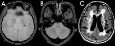
Figure 1 Brain images of the patient before admission. Three months prior to admission, T2*-weighted GRE MRI showed multiple cerebral microbleeds in the thalamus (A) with no abnormal vasculature in the medulla (B). The fluid-attenuated inversion recovery image showed diffuse periventricular white-matter changes (C).
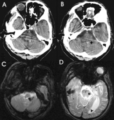
Figure 2 Brain images of the patient at admission. Brain CT (A, B) and T2*-weighted GRE MRI (C, D) showed hemorrhage in the left dorsal medulla, which ruptured the dorsal medulla and extended into the fourth ventricle (arrow). The layered blood-CSF pattern of the intraventricular hemorrhage was due to backward flow of blood into the occipital horn of the left lateral ventricle (D) (arrowhead).
Reference
-
1. Mastaglia FL, Edis B, Kakulas BA. Medullary hemorrhage: a report of two cases. J Neurol Neurosurg Psychiatry. 1969. 32:221–225.2. Kempe LG. Surgical removal of an intramedullary haematoma simulating Wallenberg\'s syndrome. J Neurol Neurosurg Psychiatry. 1964. 27:78–80.
Article3. Cohen HC, Tucker WS, Humphreys RP, Perrin RJ. Angiographically cryptic histologically verified cerebrovascular malformations. Neurosurgery. 1982. 10:704–714.
Article4. Morel-Maroger A, Metzger J, Bories J, Gardeur D, Verger JB, Noel MC. Non-lethal brain stem hematomas in hypertensive patients. Rev Neurol (Paris). 1982. 138:437–445.5. Biller J, Gentry LR, Adams HP Jr, Morris DC. Spontaneous hemorrhage in the medulla oblongata: clinical MR correlations. J Comput Assist Tomogr. 1986. 10:303–306.6. Barinagarrementeria F, Cantu C. Primary medullary hemorrhage. Report of four cases and review of the literature. Stroke. 1994. 25:1684–1687.
Article7. Neumann PE, Mehler MF, Horoupian DS. Primary medullary hypertensive hemorrhage. Neurology. 1985. 35:925–928.
Article
