Unusual Diaphragmatic Hernias Mimicking Cardiac Masses
- Affiliations
-
- 1Department of Cardiovascular Medicine, Gachon University Gil Medical Center, Incheon, Korea. heart@gachon.ac.kr
- 2Gachon Cardiovascular Research Institute, Gachon University School of Medicine, Incheon, Korea.
- KMID: 1806920
- DOI: http://doi.org/10.4250/jcu.2015.23.2.107
Abstract
- Hiatal hernia and Morgagni hernia are sorts of diaphragmatic hernias that are rarely detected on transthoracic echocardiography. Although echocardiographic findings have an important role for differential diagnosis of cardiac masses, we often might overlook diaphragmatic hernia. We report three cases of diaphragmatic hernias having specific features. The first case is huge hiatal hernia that encroaches left atrium with internal swirling flow on transthoracic echocardiography. The second case is a hiatal hernia that encroaches on both atria, incidentally detected on preoperative echocardiography. The third case is Morgagni hernia which encroaches on the right atrium only. So, we need to consider possibility of diaphragmatic hernia when we find a cardiac mass with specific echocardiographic features.
MeSH Terms
Figure
-
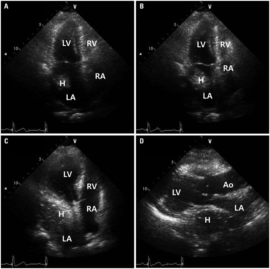
Fig. 1 Hiatal hernia with internal swirling flow. A: Apical 4-chamber view of transthoracic echocardiography shows intracardiac mass in left atrium. B: Posterior plane of (A) shows larger intracardiac mass in left atrium. C: More posterior plane of (B) shows huge extrinsic mass that compress left atrium with internal swirling flow. D: Parasternal long axis view of transthoracic echocardiography shows huge extrinsic mass that compress left atrium with internal swirling flow. Ao: aorta, H: hiatal hernia, LA: left atrium, LV: left ventricle, RA: right atrium, RV: right ventricle.
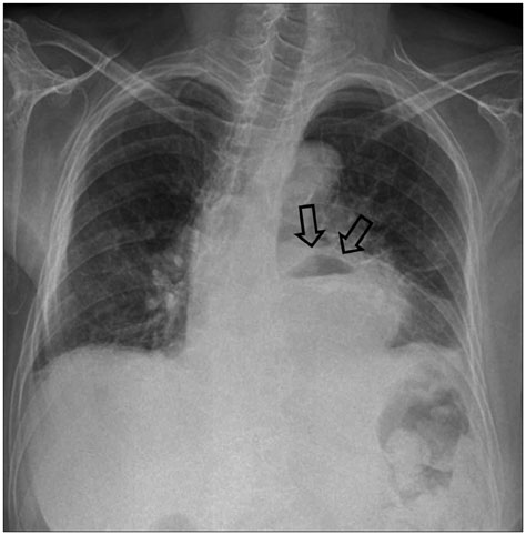
Fig. 2 Chest X-ray showing air-fluid level that suggest large hiatal hernia (arrows).
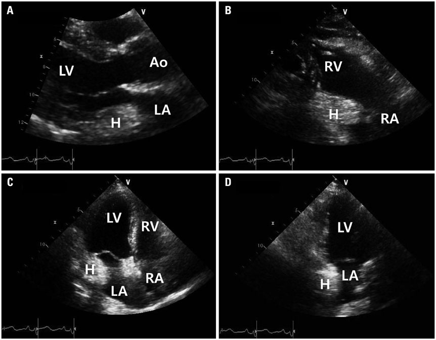
Fig. 3 Echocardiographic findings of hiatal hernia showing cardiac mass involving both atria. A: Parasternal long axis view of transthoracic echocardiography shows huge extracardiac mass (3.8 × 3.5 cm) adjacent to the left heart compressing left atrium. B: Parasternal long axis RV inflow view of transthoracic echocardiography shows mobile echogenic mass (3.4 × 2.8 cm) in right atrium. Apical 4-chamber view (C) and apical 2-chamber view (D) of transthoracic echocardiography shows mobile echogenic mass that compressing left atrium. Ao: aorta, H: hiatal hernia, LA: left atrium, LV: left ventricle, RA: right atrium, RV: right ventricle.

Fig. 4 Chest computed tomography of hiatal hernia showing cardiac mass involving both atria. Chest computed tomography shows large hiatal hernia compressing left atrium (A and B) and right atrium (C and D) (arrows).
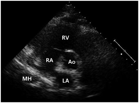
Fig. 5 Echocardiographic findings of Morgagni's hernia. Parasternal short axis view of transthoracic echocardiography shows large hypoechoic mass encroaching posterior aspect of right atrium. Ao: aorta, LA: left atrium, MH: Morgagni hernia, RA: right atrium, RV: right ventricle.

Fig. 6 Chest computed tomography of Morgagni's hernia. Chest computed tomography shows prominent fat tissue in right cardiophrenic area with suspicious internal vasculature that suggested Morgagni's hernia (arrows).
Cited by 1 articles
-
An Incidental Discovery of Morgagni Hernia in an Elderly Patient Presented with Chronic Dyspepsia
Duk Ki Kim, Hee Seok Moon, Hyeon Yong Jung, Jae Kyu Sung, Sun Hyeong Gang, Myeong Hee Kim
Korean J Gastroenterol. 2017;69(1):68-73. doi: 10.4166/kjg.2017.69.1.68.
Reference
-
1. Kim J, Han D, Sohn C, Kim JS, Park YH. Catheter ablation of ventricular arrhythmias via the radial artery in a patient with prior myocardial infarction and peripheral vascular disease. Korean Circ J. 2012; 42:632–637.
Article2. Oishi Y, Ishimoto T, Nagase N, Mori K, Fujimoto S, Hayashi S, Ochi Y, Kobayashi K, Tabata T, Oki T. Syncope upon swallowing caused by an esophageal hiatal hernia compressing the left atrium: a case report. Echocardiography. 2004; 21:61–64.
Article3. Nishimura RA, Tajik AJ, Schattenberg TT, Seward JB. Diaphragmatic hernia mimicking an atrial mass: a two-dimensional echocardiographic pitfall. J Am Coll Cardiol. 1985; 5:992–995.
Article4. D'Cruz IA, Hancock HL. Echocardiographic characteristics of diaphragmatic hiatus hernia. Am J Cardiol. 1995; 75:308–310.5. Khouzam RN, Akhtar A, Minderman D, Kaiser J, D'Cruz IA. Echocardiographic aspects of hiatal hernia: a review. J Clin Ultrasound. 2007; 35:196–203.
Article6. Torfs CP, Curry CJ, Bateson TF, Honoré LH. A population-based study of congenital diaphragmatic hernia. Teratology. 1992; 46:555–565.
Article7. Lin ST, Moss DM, Henderson SO. A case of Morgagni hernia presenting as pneumonia. J Emerg Med. 1997; 15:297–301.
Article8. Sutro WH, King SJ. Computed tomography of Morgagni hernia. N Y State J Med. 1987; 87:520–521.9. Yeager BA, Guglielmi GE, Schiebler ML, Gefter WB, Kressel HY. Magnetic resonance imaging of Morgagni hernia. Gastrointest Radiol. 1987; 12:296–298.
Article
- Full Text Links
-
- Actions
-
Cited
- CITED
-
- Close
- Share
-
- Similar articles
-
- A Case Report and Systemic Analysis of Articles for Morgagni Diaphragmatic Hernia
- Cardiac Arrest Due to Unrecognized Congenital Diaphragmatic Hernia
- Unilateral Congenital Diaphragmatic Eventration Mimicking Congenital Diaphragmatic Hernia
- A Case of Morgagni Hernia
- A Case of Maternal Diaphragmatic Hernia in Pregnancy
