J Korean Assoc Pediatr Surg.
2015 Jun;21(1):14-16. 10.13029/jkaps.2015.21.1.14.
Omphalocele with Double Prolapse of Ileum through Patent Vitellointestinal Duct: A Rare Presentation
- Affiliations
-
- 1Department of Pediatric Surgery, Postgraduate Institute of Medical Education and Research, Chandigarh, India. monikabawa@hotmail.com
- KMID: 1804839
- DOI: http://doi.org/10.13029/jkaps.2015.21.1.14
Abstract
- Although Meckel's diverticulum is the most common vitellointestinal duct (VID) anomaly, patent vitellointestinal duct (PVID) is the most common symptomatic embryological defect. Patient may present with the anomaly itself or due to complications like intestinal obstruction secondary to volvulus, intussusception or adhesions. Prolapse occurs if the diverticulum is wide-mouthed enough to allow bowel to come out or due to increased intra-abdominal pressure like cry or cough. Bowel prolapse through PVID is rare and double prolapse of proximal as well as distal loop in a newborn is extremely rare. Omphalocele with prolapsing bowel through PVID as found in our index case is even rarer in literature. The pediatric surgeon should be familiar with these varied manifestations in the newborn because the prolapsed bowel can progress to gangrene and complications if not identified and operated upon early.
MeSH Terms
Figure
-
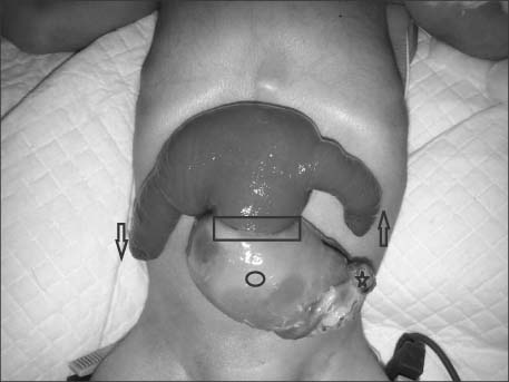
Fig. 1 Umbilicus (asterisk), omphalocele (circle), site of prolapsed (rectangle) of patent vitellointestinal duct, proximal intussuscepted ileum (upwards arrow) from which some meconium was discharging at time of examination in emergency, distal intussuscepted ileum (downwards arrow).

Fig. 2 Intraoperative picture with arrow and forceps at site of wide mouthed patent vitellointestinal duct from where intussuscepting ileal loops were reduced.
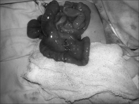
Fig. 3 Intraoperative picture after resection and ileoileal end to end anastomosis at site of prolapsed patent vitellointestinal duct.
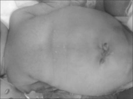
Fig. 4 Postoperative picture shows well healed site of incision and umbilicoplasty.
Reference
-
1. Cilley RE. Disorders of the umbilicus. In : Grosfeld JL, O'Neill JA, Coran AG, Fonkalsrud EW, editors. Pediatric surgery. 6th ed. St. Louis: Mosby/Elsevier;2006. p. 1143–1156.2. Chiang LS. Vitelline duct remnant appearing as a hemorrhagic umbilical mass. JAMA. 1982; 247:2812–2813.3. Agrawal S, Memon A. Patent vitellointestinal duct. BMJ Case Rep. 2010; doi: 10.1136/bcr.12.2009.2594.4. Ameh EA, Mshelbwala PM, Dauda MM, Sabiu L, Nmadu PT. Symptomatic vitelline duct anomalies in children. S Afr J Surg. 2005; 43:84–85.5. Soutar SF, Douglas DM, Dennison WM. Patent vitello-intestinal duct; the risk of obstruction due to prolapse. Br J Surg. 1958; 45:617–620.6. Borkar N. A prolapsing vitello-intestinal duct in newborn. APSP J Case Rep. 2013; 4:32.7. Davis RM, Kehm RW. Omphalocele with patent vitellointestinal duct and ileal prolapse. Am J Surg. 1967; 113:571–573.8. Rohatgi M, Gorthi SN. Omphalocele with patent omphalomesenteric duct and ileal prolapse. Indian J Pediatr. 1984; 51:119–123.9. Storms P, Pexsters J, Vandekerkhof J. Small omphalocele with ileal prolapse through a patent omphalomesenteric duct. A case report and review of literature. Acta Chir Belg. 1988; 88:392–394.10. Panait N, Michel F, D'Ercole C, Merrot T. Esophageal atresia, small omphalocele and ileal prolapse through a patent omphalomesenteric duct: a methamizole embryopathy? J Pediatr Surg. 2013; 48:E9–E11.11. Blair SP, Beasley SW. Intussusception of vitello-intestinal tract through an exomphalos in trisomy 13. Ped Surg Int. 1989; 4:422–423.12. Mohite PN, Bhatnagar AM, Hathila VP, Mistry JH. Patent vitellointestinal duct with prolapse of inverted loop of small intestine: a case report. J Med Case Rep. 2007; 1:49.
