Calcified Carcinoma of the Gallbladder with Calcified Nodal Metastasis Presenting as a Porcelain Gallbladder: A Case Report
- Affiliations
-
- 1Department of Radiology, Kangdong Sacred Heart Hospital, Hallym University College of Medicine, Seoul, Korea.
- 2Department of Internal Medicine, Kangdong Sacred Heart Hospital, Hallym University College of Medicine, Seoul, Korea.
- 3Department of Pathology, Kangdong Sacred Heart Hospital, Hallym University College of Medicine, Seoul, Korea. esnam2003@yahoo.co.kr
Abstract
- Porcelain gallbladder is regarded as a risk factor of gallbladder cancer. A porcelain gallbladder with calcified regional lymph nodes was found using computed tomography (CT) and magnetic resonance imaging (MRI) in a 43-year-old man who presented with nausea, vomiting, and abdominal pain. His cholecystectomy specimen showed diffuse wall thickening and contained small gallstones. Histological examination revealed diffuse infiltrative adenocarcinoma with extensive intratumoral calcification (calcified carcinoma). The majority of the calcified material was located within or replaced the tumor glands, and was not found in the stroma. A lymph node was totally replaced with a calcified metastatic adenocarcinoma. To the best of our knowledge, only one case of calcified lymph node metastasis from a calcified carcinoma of the gallbladder has been previously reported in the literature. We herein add a case of calcified carcinoma of the gallbladder with calcified lymph node metastasis, presenting as a porcelain gallbladder on CT and MRI.
Keyword
MeSH Terms
Figure
-
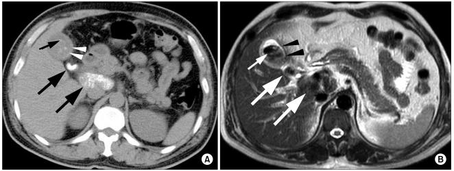
Fig. 1 (A) Unenhanced computed tomography scan showed circumferential calcification in the thickened wall of the gallbladder (arrowheads) and extensive calcified regional lymph nodes (arrows). Multiple punctuated hyperdense lesions, which were found to be gallstones, were also noted inside the gallbladder lumen (small arrow). (B) T2-weighted magnetic resonance image at 9000/1200 (TR/TE) demonstrated areas of signal void in the gallbladder wall (arrowheads), within enlarged lymph nodes (arrows), and inside the gallbladder lumen (small arrow).
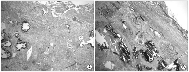
Fig. 2 (A) Histological examination of the resected gallbladder revealed denuded mucosa and infiltrative, well-differentiated adenocarcinoma with numerous fine to coarse calcifications. The majority of the calcified material was located within or replaced the tumor glands, and not found in the stroma (H&E, ×40). (B) Histological examination of the regional lymph node revealed metastatic adenocarcinoma with calcification (H&E, ×40).
Reference
-
1. Osler W. The principles and practice of medicine. 1925. 10th ed. New York: Appleton;p. 574.2. Polk HC Jr. Carcinoma and the calcified gall bladder. Gastroenterology. 1966; 50:582–585. PMID: 4286335.
Article3. Ashur H, Siegal B, Oland Y, Adam YG. Calcified ballbladder (porcelain gallbladder). Arch Surg. 1978; 113:594–596. PMID: 646619.4. Towfigh S, McFadden DW, Cortina GR, Thompson JE Jr, Tompkins RK, Chandler C, et al. Porcelain gallbladder is not associated with gallbladder carcinoma. Am Surg. 2001; 67:7–10. PMID: 11206901.5. Stephen AE, Berger DL. Carcinoma in the porcelain gallbladder: a relationship revisited. Surgery. 2001; 129:699–703. PMID: 11391368.
Article6. Shimizu M, Miura J, Tanaka T, Itoh H, Saitoh Y. Porcelain gallbladder: relation between its type by ultrasound and incidence of cancer. J Clin Gastroenterol. 1989; 11:471–476. PMID: 2668401.7. Kwon AH, Inui H, Matsui Y, Uchida Y, Hukui J, Kamiyama Y. Laparoscopic cholecystectomy in patients with porcelain gallbladder based on the preoperative ultrasound findings. Hepatogastroenterology. 2004; 51:950–953. PMID: 15239221.8. Goetze TO, Paolucci V. Adequate extent in radical re-resection of incidental gallbladder carcinoma: analysis of the German Registry. Surg Endosc. 2010; 24:2156–2164. PMID: 20177938.
Article9. Berk RN, Armbuster TG, Saltzstein SL. Carcinoma in the porcelain gallbladder. Radiology. 1973; 106:29–31. PMID: 4682728.
Article10. Joo YE, Kim HS, Choi SK, Rew JS, Kim HJ, Kang HK, et al. Case of mucinous adenocarcinoma with porcelain gallbladder. J Gastroenterol Hepatol. 2003; 18:995–998. PMID: 12859733.
Article11. Sweeney DJ, Low VH, Robbins PD, Yu SF. Calcified lymph node metastases in adenocarcinoma of the colon. Australas Radiol. 1994; 38:233–234. PMID: 7945124.
Article12. Batlan LE. Calcification within the stomach wall in gastric malignancy: case report and review of literature. Am J Roentgenol Radium Ther Nucl Med. 1954; 72:788–794.13. Nagakura S, Shirai Y, Yamai K, Hatakeyama K. Calcification in mucinous cholangiocellular carcinoma. Hepatogastroenterology. 1999; 46:465–466. PMID: 10228844.14. Parker GW, Joffe N. Calcifying primary mucus-producing adenocarcinoma of the gallbladder. Br J Radiol. 1972; 45:468–469. PMID: 5029033.
Article15. Rogers LF, Lastra MP, Lin KT, Bennett D. Calcifying mucinous adenocarcinoma of the gallbladder. Am J Gastroenterol. 1973; 59:441–445. PMID: 4350100.
