Primary Gastric Choriocarcinoma: Two Case Reports and Review of the Literatures
- Affiliations
-
- 1Department of Internal Medicine, Korea Cancer Center Hospital, Korea Institute of Radiological and Medical Sciences, Seoul, Korea. hyejin@kcch.re.kr
- 2Department of Pathology, Korea Cancer Center Hospital, Korea Institute of Radiological and Medical Sciences, Seoul, Korea.
- 3Department of General Surgery, Korea Cancer Center Hospital, Korea Institute of Radiological and Medical Sciences, Seoul, Korea.
Abstract
- Primary gastric choriocarcinoma (PGC) is a rare tumor, and its pathogenesis is still uncertain. Most PGCs have been reported to possess an adenocarcinoma component of variable extent, and pure PGC is especially rare. The diagnosis of PGC is confirmed by exhibition of choriocarcinomatous components on biopsy and exhibition of beta-hCG positive cell on immunohistochemical stain and elevation of the serum beta-hCG. Moreover it must be confirmed that no other site including gonads displays any tumor masses. The PGC tends to be more invasive and to have early metastasis. The median survival is known to be less than several months. We report two cases. The first case was a 62 year-old man who was diagnosed as advanced gastric cancer (AGC) by endoscopic biopsy with hepatic metasasis and received palliative chemotherapy with modified FOLFOX regimen and Genexol plus cisplatin regimen. He underwent subtotal gastrectomy due to perforation of the stomach during chemotherapy. On post-operative biopsy, He wasre-diagnosed as PGC and received another palliative chemotherapy modified FOLFIRI, BEP, EMACO, VIP. However, multiple liver metastases were aggravated, and also serum AFP level increased. Ultimately, the paient died 10 months after initial diagnosis. Another case was a 45 year-old man. On endoscopic biopsy, he was diagnosed as AGC of adenocarcinoma. On Chest and Abdomen CT, multiple pulmonary and hepatic metastasis were also confirmed. On liver biopsy, He was diagnosed as PGC. The immunohistochemical stains were performed and the results were cytokeratin positive, EMA negative and beta-hCG weak positive. The serum beta-hCG level was highly elevated. BEP, VIP and EMA/CO combination therapy were administered, but he died at 12th months after the initial diagnosis.
Keyword
MeSH Terms
-
Abdomen
Adenocarcinoma
Antineoplastic Combined Chemotherapy Protocols
Biopsy
Choriocarcinoma
Cisplatin
Coloring Agents
Female
Fluorouracil
Gastrectomy
Gonads
Keratins
Leucovorin
Liver
Neoplasm Metastasis
Organoplatinum Compounds
Pregnancy
Stomach
Stomach Neoplasms
Thorax
Antineoplastic Combined Chemotherapy Protocols
Cisplatin
Coloring Agents
Fluorouracil
Keratins
Leucovorin
Organoplatinum Compounds
Figure
-

Fig. 1 Endoscopic finding is shows a huge ulceroinfiltrative mucosal lesion on antrum of stomach.
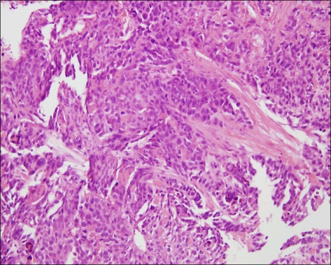
Fig. 2 Stomach. Microscopic finding of stomach shows moderately differentiated adenocarcinoma. (H&E, ×200).

Fig. 3 Abdominal CT. Abdominal computed tomographic finding shows low attenuating mass and nodules in both hepatic lobes with metastatic lymphadenopathies.
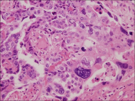
Fig. 4 Stomach. Microscopic finding of stomach shows choriocarcinoma, characterized by dimorhpic plexiform pattern (H&E, ×400).
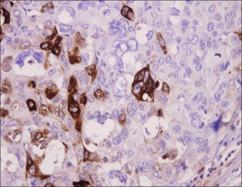
Fig. 5 Stomach. β-hCG stain. Immunohistochemical staining for β-hCG of stomach shows a positive reaction (peroxidase-HRP, ×400).

Fig. 6 Liver. Microscopic finding of liver shows the hepatic infiltration of choriocarcinoma (H&E, ×100)
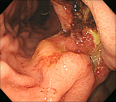
Fig. 7 Gastric endoscopic feature shows a huge ulceroinfiltrative mucosal lesion on lesser curvature of the body.

Fig. 8 Abdominal CT. Two huge and several smaller variable sized low density lesions are seen in the liver.
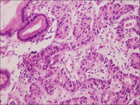
Fig. 9 Stomach. Microscopic finding of stomach shows moderately differentiated adenocarcinoma (H&E, ×400).

Fig. 10 Liver. Microscopic finding of liver shows the hepatic infiltration of choriocarcinoma. Some of the multinucleated giant cells (arrows), consistent with syncytiorophoblast (H&E, ×400).
Reference
-
1. Liu Z, Mira JL, Cruz-Caudillo JC. Primary gastric chorio carcinoma: a case report and review of the literature. Arch Pathol Lab Med. 2001; 125:1601–1604. PMID: 11735700.2. Kameya T, Kuramoto H, Suzuki K, Kenjo T, Oshikiri T, Hayashi H, et al. A human gastric choriocarcinoma cell line with human chorionic gonadotropin and placental alkaline phosphatase production. Cancer Res. 1975; 35:2025–2032. PMID: 1170940.3. Kobayashi A, Hasebe T, Endo Y, Sasaki S, Konishi M, Sugito M, et al. Primary gastric choriocarcinoma: two case reports and a pooled analysis of 53 cases. Gastric Cancer. 2005; 8:178–185. PMID: 16086121.
Article4. Nam SH, Im SA, Bae KS, Kang KS, Kang IS, Kwon JM, et al. A case of primary gastric choriocarcinoma presenting with amenorrhea. Cancer Res Treat. 2002; 34:457–460.
Article5. Noguchi T, Takeno S, Sato T, Takahashi Y, Uchida Y, Yokoyama S. A patient with primary gastric choriocarcinoma who received a correct preoperative diagnosis and achieved prolonged survival. Gastric Cancer. 2002; 5:112–117. PMID: 12111588.
Article6. Ozaki H, Ito I, Sano R, Hirota T, Shimosato Y. A case of choriocarcinoma of the stomach. Jpn J Clin Oncol. 1971; 1:83.7. Koritschoner R. Uber ein chorioepithelium ohne primartumor mit abnormalanger Latenzzeit. Beitr Z Path Anat. 1920; 66:501.8. Pick L. Uber die chorioepthelahnlich metastasierende from des magencarcinomas. Klin Wochenscher. 1926; 5:1728.9. Hartz PH, Ramirez CA. Coexistence of carcinoma and chorioepithelioma in the stomach of young man. Cancer. 1953; 6:319–326. PMID: 13032923.10. Liu AY, Chan WY, Ng EK, Zhang X, Li BC, Chow JH, et al. Gastric choriocarcinoma shows characteristics of adenocarcinoma and gestational choriocarcinoma: a comparative genomic hybridization and fluorescence in situ hybridization study. Diagn Mol Pathol. 2001; 10:161–165. PMID: 11552718.
Article11. Jung KC, Kim WH, Kim YI, Choe KJ. Gastric adenocarcinoma with choriocarcinomatous and hepatoid differentiation: report of a case. Korean J Pathol. 1994; 28:409–413.12. Kawashima Y, Ishikawa H, Hada M, Sakata K, Hirai T, Asaumi S, et al. A case of primary gastric choriocarcinoma. Gan No Rinsho. 1989; 35:1466–1472. PMID: 2681880.
