Ultrasound Follow-Up of Testicular Adrenal Rest Tumors with Congenital Adrenal Hyperplasia: Report of Three Cases
- Affiliations
-
- 1Department of Radiology, Dong-A University Hospital, Busan, Korea. jedidw@naver.com
- KMID: 1801557
- DOI: http://doi.org/10.3348/jksr.2014.71.6.324
Abstract
- While testicular adrenal rest tumor is generally a rare intratesticular tumor, it is frequent in patients with congenital adrenal hyperplasia. The tumors are diagnosed and followed up by ultrasound examination because these tumors are non-palpable and symptomless in most cases and always benign. Ultrasound imaging features change depending on how congenital adrenal hyperplasia is controlled. We herein report three cases of testicular adrenal rest tumors with different usual and unusual imaging findings and follow-up imaging. Patient 1 was a 14-year-old boy who presented with poor compliance to medication. Patient 2 and 3 were a 10-year-old and 13-year-old boy who presented with precocious puberty and short stature, respectively. Ultrasound examinations demonstrated oval hypoechoic masses and irregular speculated hyperechoic masses in the testes and different serial imaging findings.
MeSH Terms
Figure
-
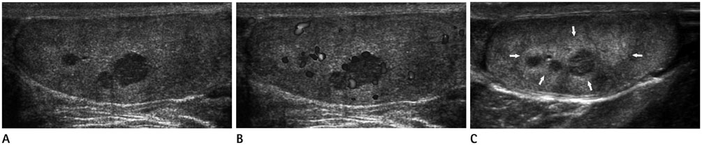
Fig. 1 Ultrasound (US) images of testicular adrenal rest tumors in a 14-year-old boy. A. Gray-scale US shows three well defined oval hypoechoic masses with no posterior acoustic shadowing around the mediastinum of the left testis. B. Color Doppler US shows nodular vascularity in peripheral portion of the masses. C. On US performed 4 years later, the masses decrease in size. However, ill-defined hyperechoic lesions (arrows) surrounding the masses develop.

Fig. 2 Ultrasound (US) images of testicular adrenal rest tumors in a 10-year-old boy. A. Gray-scale US shows a small well defined oval hypoechoic mass with no posterior acoustic shadowing in the right mediastinum testis. B. Follow-up US 6 months later shows complete resolution of the previous mass. C. Follow-up color Doppler US 23 months later shows newly well defined oval hypoechoic masses with nodular vascularity developed in the mediastinum testis.

Fig. 3 Ultrasound (US) images of testicular adrenal rest tumors in a 13-year-old boy. A. Gray-scale US shows an irregular speculated hyperechoic mass (arrows) around the mediastinum testis without posterior acoustic shadowing and small oval well defined hypoechoic mass (arrowheads) adjacent to main mass. B. Color Doppler US shows increased nodular vascularity in the mass. C. Follow-up US 6 months later shows no change in the size and shape of the main mass, but complete resolution of the small masses adjacent to main mass.
Reference
-
1. Claahsen-van der Grinten HL, Otten BJ, Stikkelbroeck MM, Sweep FC, Hermus AR. Testicular adrenal rest tumours in congenital adrenal hyperplasia. Best Pract Res Clin Endocrinol Metab. 2009; 23:209–220.2. Stikkelbroeck NM, Suliman HM, Otten BJ, Hermus AR, Blickman JG, Jager GJ. Testicular adrenal rest tumours in postpubertal males with congenital adrenal hyperplasia: sonographic and MR features. Eur Radiol. 2003; 13:1597–1603.3. Delfino M, Elia J, Imbrogno N, Argese N, Mazzilli R, Toscano V, et al. Testicular adrenal rest tumors in patients with congenital adrenal hyperplasia: prevalence and sonographic, hormonal, and seminal characteristics. J Ultrasound Med. 2012; 31:383–388.4. Avila NA, Shawker TS, Jones JV, Cutler GB Jr, Merke DP. Testicular adrenal rest tissue in congenital adrenal hyperplasia: serial sonographic and clinical findings. AJR Am J Roentgenol. 1999; 172:1235–1238.5. Clark RV, Albertson BD, Munabi A, Cassorla F, Aguilera G, Warren DW, et al. Steroidogenic enzyme activities, morphology, and receptor studies of a testicular adrenal rest in a patient with congenital adrenal hyperplasia. J Clin Endocrinol Metab. 1990; 70:1408–1413.6. Wilkins L, Fleishmann W, Howard JE. Macrogenitosomia precox associated with hyperplasia of the androgenic tissue of the adrenal and death from corticoadrenal insufficiency. Endocrinology. 1940; 26:385–395.7. Vanzulli A, DelMaschio A, Paesano P, Braggion F, Livieri C, Angeli E, et al. Testicular masses in association with adrenogenital syndrome: US findings. Radiology. 1992; 183:425–429.8. Chrousos GP, Loriaux DL, Sherins RJ, Cutler GB Jr. Unilateral testicular enlargement resulting from inapparent 21-hydroxylase deficiency. J Urol. 1981; 126:127–128.9. Dogra VS, Gottlieb RH, Oka M, Rubens DJ. Sonography of the scrotum. Radiology. 2003; 227:18–36.10. Nagamine WH, Mehta SV, Vade A. Testicular adrenal rest tumors in a patient with congenital adrenal hyperplasia: sonographic and magnetic resonance imaging findings. J Ultrasound Med. 2005; 24:1717–1720.11. Seidenwurm D, Smathers RL, Kan P, Hoffman A. Intratesticular adrenal rests diagnosed by ultrasound. Radiology. 1985; 155:479–481.
- Full Text Links
-
- Actions
-
Cited
- CITED
-
- Close
- Share
-
- Similar articles
-
- A case of testicular adrenal rest tumor in a male child with congenital adrenal hyperplasia
- Testicular Adrenal Rest Tumor in 11-Beta-Hydroxylase Deficiency Driven Congenital Adrenal Hyperplasia
- Erratum: Ultrasound Follow-Up of Testicular Adrenal Rest Tumors with Congenital Adrenal Hyperplasia: Report of Three Cases
- A Case of Congenital Adrenal Hyperplasia Combined with a Testicular Adrenal Rest Tumor and Adrenal Incidentaloma
- Testicular adrenal rest tumors in a patient with untreated congenital adrenal hyperplasia
