Clin Endosc.
2015 Mar;48(2):142-146. 10.5946/ce.2015.48.2.142.
Light-Emitting Diode-Assisted Narrow Band Imaging Video Endoscopy System in Head and Neck Cancer
- Affiliations
-
- 1Department of Otolaryngology, Head and Neck Surgery, Cathay General Hospital, Taipei, Taiwan. drtony@seed.net.tw
- 2Electronic and Optoelectronic Research Laboratories, Industrial Technology Research Institute, Hsinchu, Taiwan.
- KMID: 1801205
- DOI: http://doi.org/10.5946/ce.2015.48.2.142
Abstract
- BACKGROUND/AIMS
To validate the effectiveness of a newly developed light-emitting diode (LED)-narrow band imaging (NBI) system for detecting early malignant tumors in the oral cavity.
METHODS
Six men (mean age, 51.5 years) with early oral mucosa lesions were screened using both the conventional white light and LED-NBI systems.
RESULTS
Small elevated or ulcerative lesions were found under the white light view, and typical scattered brown spots were identified after shifting to the LED-NBI view for all six patients. Histopathological examination confirmed squamous cell carcinoma. The clinical stage was early malignant lesions (T1), and the patients underwent wide excision for primary cancer. This is the pilot study documenting the utility of a new LED-NBI system as an adjunctive technique to detect early oral cancer using the diagnostic criterion of the presence of typical scattered brown spots in six high-risk patients.
CONCLUSIONS
Although large-scale screening programs should be established to further verify the accuracy of this technology, its lower power consumption, lower heat emission, and higher luminous efficiency appear promising for future clinical applications.
MeSH Terms
Figure
-
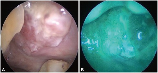
Fig. 1 Case 6, (A) left buccal mucosa lesion (1.8×1.7 cm2). (B) The narrow band imaging examination uncovered a well-demarcated brownish area with scattered brown spots. Histopathology revealed squamous cell carcinoma, moderately differentiated.
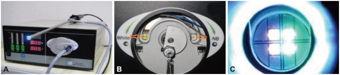
Fig. 2 (A) Light-emitting diode (LED)-narrow band imaging light source PR650, cursors for 0% to 100% white, green/blue light mix. (B) Left ⇨: white LED chip; right ⇦: green/blue LED chips. (C) Close-up view: green/blue LED chips.
Reference
-
1. Lin YC, Wang WH, Lee KF, Tsai WC, Weng HH. Value of narrow band imaging endoscopy in early mucosal head and neck cancer. Head Neck. 2012; 34:1574–1579. PMID: 22290602.
Article2. Forastiere A, Koch W, Trotti A, Sidransky D. Head and neck cancer. N Engl J Med. 2001; 345:1890–1900. PMID: 11756581.
Article3. Muto M, Nakane M, Katada C, et al. Squamous cell carcinoma in situ at oropharyngeal and hypopharyngeal mucosal sites. Cancer. 2004; 101:1375–1381. PMID: 15368325.
Article4. Piazza C, Dessouky O, Peretti G, Cocco D, De Benedetto L, Nicolai P. Narrow-band imaging: a new tool for evaluation of head and neck squamous cell carcinomas. Review of the literature. Acta Otorhinolaryngol Ital. 2008; 28:49–54. PMID: 18669067.5. Gono K, Yamazaki K, Doguchi N, et al. Endoscopic observation of tissue by narrowband illumination. Opt Rev. 2003; 10:211–215.
Article6. Wang WH, Lin YC, Lee KF, Weng HH. Nasopharyngeal carcinoma detected by narrow-band imaging endoscopy. Oral Oncol. 2011; 47:736–741. PMID: 21393053.
Article7. Katada C, Nakayama M, Tanabe S, et al. Narrow band imaging for detecting superficial oral squamous cell carcinoma: a report of two cases. Laryngoscope. 2007; 117:1596–1599. PMID: 17597626.
Article8. Watanabe A, Taniguchi M, Tsujie H, Hosokawa M, Fujita M, Sasaki S. The value of narrow band imaging for early detection of laryngeal cancer. Eur Arch Otorhinolaryngol. 2009; 266:1017–1023. PMID: 18982341.
Article9. Sano Y, Kobayashi M, Hamamoto Y, et al. New diagnostic method based on color imaging using narrowband imaging (NBI) endoscopy system for gastrointestinal tract. Gastrointest Endosc. 2001; 53:AB125.10. Lin YC, Watanabe A, Chen WC, Lee KF, Lee IL, Wang WH. Narrowband imaging for early detection of malignant tumors and radiation effect after treatment of head and neck cancer. Arch Otolaryngol Head Neck Surg. 2010; 136:234–239. PMID: 20231639.
Article11. Wang WH, Lin YC, Chen WC, Chen MF, Chen CC, Lee KF. Detection of mucosal recurrent nasopharyngeal carcinomas after radiotherapy with narrow-band imaging endoscopy. Int J Radiat Oncol Biol Phys. 2012; 83:1213–1219. PMID: 22099040.
Article12. Jo WK, Eun SS, Shin SH. Feasibility of light-emitting diode uses for annular reactor inner-coated with TiO2 or nitrogen-doped TiO2 for control of dimethyl sulfide. Photochem Photobiol. 2011; 87:1016–1023. PMID: 21707635.
Article13. Epstein JB, Silverman S Jr, Epstein JD, Lonky SA, Bride MA. Analysis of oral lesion biopsies identified and evaluated by visual examination, chemiluminescence and toluidine blue. Oral Oncol. 2008; 44:538–544. PMID: 17996486.
Article14. Sciubba JJ. Oral cancer and its detection. History-taking and the diagnostic phase of management. J Am Dent Assoc. 2001; 132(Suppl):12S–18S. PMID: 11803647.
- Full Text Links
-
- Actions
-
Cited
- CITED
-
- Close
- Share
-
- Similar articles
-
- Interobserver Agreement in Using Magnifying Narrow Band Imaging System
- Clinical Role of Magnifying Endoscopy with Narrow-band Imaging in the Diagnosis of Early Gastric Cancer
- Usefulness of Narrow-Band Imaging in Endoscopic Submucosal Dissection of the Stomach
- Clinical Application of Magnifying Endoscopy with Narrow-Band Imaging in the Stomach
- The shear bond strength and adhesive failure pattern in bracket bonding with different light-curing methods
