The Added Value of Double Dose Gadolinium Enhanced 3D T2 Fluid-Attenuated Inversion Recovery for Evaluating Small Brain Metastases
- Affiliations
-
- 1Department of Radiology, Yonsei University College of Medicine, Seoul, Korea. slee@yuhs.ac
- 2Department of Neurosurgery, Yonsei University College of Medicine, Seoul, Korea.
- KMID: 1799485
- DOI: http://doi.org/10.3349/ymj.2014.55.5.1231
Abstract
- PURPOSE
Single dose gadolinium (Gd) enhanced fluid-attenuated inversion recovery (FLAIR) is helpful for visualizing superficial parenchymal metastases. However, the usefulness of FLAIR with a higher dose of Gd is uncertain. The aim of our study was two-folds: first, to prove that the signal to noise ratio (SNR) of small brain metastases is higher than large brain metastases on double-dose (DD) enhanced FLAIR and, second, to explore the added value of DD Gd enhanced FLAIR in relation to T1 GRE for evaluating small brain metastases.
MATERIALS AND METHODS
For the first purpose, 50 pairs of small (2 mm
RESULTS
The SNR and CR of small brain metastases were significantly higher than those of large brain metastases (p<0.001). In qualitative analysis, the diagnostic sensitivities for small brain metastases were significantly higher for 3D T1 GRE plus 3D T2 FLAIR than 3D T1 GRE alone regardless of scan time (p<0.001).
CONCLUSION
Small brain metastases showed higher signal intensity than large brain metastases on the DD Gd enhanced 3D T2 FLAIR images. DD Gd enhanced 3D T2 FLAIR imaging may have a complementary role to 3D T1 GRE for evaluating small brain metastases.
Keyword
MeSH Terms
Figure
-
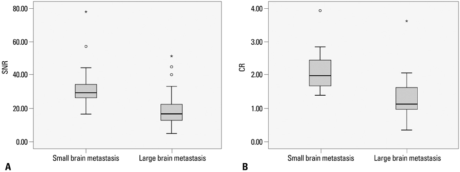
Fig. 1 Comparison of SNR and CR between small metastasis (2 mm
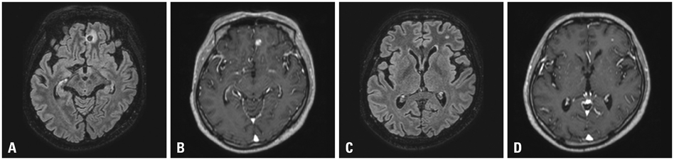
Fig. 2 A 45-year-old patient with renal cell cancer. (A) A larger brain metastasis (8 mm) with dark signal intensity and peri-lesional edema in the left frontal lobe on DD Gd-enhanced 3D T2 FLAIR (SNR: 9.61). (B) The same lesion with strong enhancement on post DD gadolinium enhanced 3D T1 GRE. (C) Another small brain metastasis (4.2 mm) in the left frontal lobe with high signal intensity on DD Gd-enhanced 3D T2 FLAIR (SNR: 21.63). (D) The same lesion with enhancement on DD Gd-enhanced 3D T1 GRE. SNR, signal-to-noise ratio; DD, double dose; Gd, gadolinium; FLAIR, fluid-attenuated inversion recovery; GRE, gradient echo.

Fig. 3 68-year-old female patient with lung cancer. (A) In first review, both reviewers missed the left frontal small brain metastases on early phase DD Gd enhanced 3D T1 GRE, because they regarded them as enhancement of the cortical vein. (B) On late phase DD Gd enhanced 3D T2 FLAIR, the same lesion with high signal intensity was more prominent and distinct from the surrounding normal parenchyma. Both reviewers retrospectively detected the left frontal lesion on 3D T1 GRE after reviewing the 3D T2 FLAIR. DD, double dose; Gd, gadolinium; FLAIR, fluid-attenuated inversion recovery; GRE, gradient echo.

Fig. 4 A 49-year-old male patient with renal cell cancer. (A) On early DD Gd-enhanced 3D T1 GRE, brain metastasis is not detected. (B) On late phase DD Gd enhanced 3D T2 FLAIR, small enhancing metastasis is clearly demonstrated in the right frontal lobe (arrow). (C) Two month follow up 3D T1 GRE reveals the small brain metastases in the right frontal lobe (dashed arrow). DD, double dose; Gd, gadolinium; FLAIR, fluid-attenuated inversion recovery; GRE, gradient echo.
Cited by 2 articles
-
Comparison of Contrast-Enhanced T2 FLAIR and 3D T1 Black-Blood Fast Spin-Echo for Detection of Leptomeningeal Metastases
Yae Won Park, Sung Jun Ahn
Investig Magn Reson Imaging. 2018;22(2):86-93. doi: 10.13104/imri.2018.22.2.86.Does Multiphasic Contrast Enhanced Fluid Attenuated Inversion Recovery Magnetic Resonance Imaging Enhance the Detectability of Small Intracerebral Metastases?
Jung Hwan Kim, Kyung Sik Yi, Chi-Hoon Choi, Seung Tae Woo, Sang-Hoon Cha
J Korean Soc Radiol. 2018;78(3):179-189. doi: 10.3348/jksr.2018.78.3.179.
Reference
-
1. Vecht CJ, Haaxma-Reiche H, Noordijk EM, Padberg GW, Voormolen JH, Hoekstra FH, et al. Treatment of single brain metastasis: radiotherapy alone or combined with neurosurgery? Ann Neurol. 1993; 33:583–590.
Article2. Patchell RA. The management of brain metastases. Cancer Treat Rev. 2003; 29:533–540.
Article3. Chang SD, Lee E, Sakamoto GT, Brown NP, Adler JR Jr. Stereotactic radiosurgery in patients with multiple brain metastases. Neurosurg Focus. 2000; 9:e3.
Article4. Yuh WT, Tali ET, Nguyen HD, Simonson TM, Mayr NA, Fisher DJ. The effect of contrast dose, imaging time, and lesion size in the MR detection of intracerebral metastasis. AJNR Am J Neuroradiol. 1995; 16:373–380.5. Sze G, Johnson C, Kawamura Y, Goldberg SN, Lange R, Friedland RJ, et al. Comparison of single- and triple-dose contrast material in the MR screening of brain metastases. AJNR Am J Neuroradiol. 1998; 19:821–828.6. Yuh WT, Engelken JD, Muhonen MG, Mayr NA, Fisher DJ, Ehrhardt JC. Experience with high-dose gadolinium MR imaging in the evaluation of brain metastases. AJNR Am J Neuroradiol. 1992; 13:335–345.7. Vogl TJ, Friebe CE, Balzer T, Mack MG, Steiner S, Schedel H, et al. [Diagnosis of cerebral metastasis with standard dose gadobutrol vs. a high dose protocol. Intraindividual evaluation of a phase II high dose study]. Radiologe. 1995; 35:508–516.8. Mathews VP, Caldemeyer KS, Lowe MJ, Greenspan SL, Weber DM, Ulmer JL. Brain: gadolinium-enhanced fast fluid-attenuated inversion-recovery MR imaging. Radiology. 1999; 211:257–263.
Article9. Terae S, Yoshida D, Kudo K, Tha KK, Fujino M, Miyasaka K. Contrast-enhanced FLAIR imaging in combination with pre- and postcontrast magnetization transfer T1-weighted imaging: usefulness in the evaluation of brain metastases. J Magn Reson Imaging. 2007; 25:479–487.
Article10. Fukuoka H, Hirai T, Okuda T, Shigematsu Y, Sasao A, Kimura E, et al. Comparison of the added value of contrast-enhanced 3D fluid-attenuated inversion recovery and magnetization-prepared rapid acquisition of gradient echo sequences in relation to conventional postcontrast T1-weighted images for the evaluation of leptomeningeal diseases at 3T. AJNR Am J Neuroradiol. 2010; 31:868–873.
Article11. Tomura N, Narita K, Takahashi S, Otani T, Sakuma I, Yasuda K, et al. Contrast-enhanced multi-shot echo-planar FLAIR in the depiction of metastatic tumors of the brain: comparison with contrast-enhanced spin-echo T1-weighted imaging. Acta Radiol. 2007; 48:1032–1037.
Article12. Davis PL, Parker DL, Nelson JA, Gillen JS, Runge VM. Interactions of paramagnetic contrast agents and the spin echo pulse sequence. Invest Radiol. 1988; 23:381–388.
Article13. Dietrich O, Raya JG, Reeder SB, Reiser MF, Schoenberg SO. Measurement of signal-to-noise ratios in MR images: influence of multichannel coils, parallel imaging, and reconstruction filters. J Magn Reson Imaging. 2007; 26:375–385.
Article14. Heverhagen JT. Noise measurement and estimation in MR imaging experiments. Radiology. 2007; 245:638–639.
Article15. Sze G, Milano E, Johnson C, Heier L. Detection of brain metastases: comparison of contrast-enhanced MR with unenhanced MR and enhanced CT. AJNR Am J Neuroradiol. 1990; 11:785–791.16. Lorusso G, Rüegg C. The tumor microenvironment and its contribution to tumor evolution toward metastasis. Histochem Cell Biol. 2008; 130:1091–1103.
Article17. Jung S, Moon KS, Jung TY, Kim IY, Lee YH, Rhu HH, et al. Possible pathophysiological role of vascular endothelial growth factor (VEGF) and matrix metalloproteinases (MMPs) in metastatic brain tumor-associated intracerebral hemorrhage. J Neurooncol. 2006; 76:257–263.
Article18. Carrier DA, Mawad ME, Kirkpatrick JB, Schmid MF. Metastatic adenocarcinoma to the brain: MR with pathologic correlation. AJNR Am J Neuroradiol. 1994; 15:155–159.19. Hinshaw DB Jr, Inouye CT. Metastatic brain neoplasms. Top Magn Reson Imaging. 1989; 1:69–78.
Article20. Chang EL, Hassenbusch SJ 3rd, Shiu AS, Lang FF, Allen PK, Sawaya R, et al. The role of tumor size in the radiosurgical management of patients with ambiguous brain metastases. Neurosurgery. 2003; 53:272–280.
Article21. Ranjan T, Abrey LE. Current management of metastatic brain disease. Neurotherapeutics. 2009; 6:598–603.
Article22. Essig M, Knopp MV, Schoenberg SO, Hawighorst H, Wenz F, Debus J, et al. Cerebral gliomas and metastases: assessment with contrast-enhanced fast fluid-attenuated inversion-recovery MR imaging. Radiology. 1999; 210:551–557.
Article23. Ercan N, Gultekin S, Celik H, Tali TE, Oner YA, Erbas G. Diagnostic value of contrast-enhanced fluid-attenuated inversion recovery MR imaging of intracranial metastases. AJNR Am J Neuroradiol. 2004; 25:761–765.24. Thomsen HS. European Society of Urogenital Radiology (ESUR). ESUR guideline: gadolinium-based contrast media and nephrogenic systemic fibrosis. Eur Radiol. 2007; 17:2692–2696.
Article
- Full Text Links
-
- Actions
-
Cited
- CITED
-
- Close
- Share
-
- Similar articles
-
- Comparison of Contrast-Enhanced T2 FLAIR and 3D T1 Black-Blood Fast Spin-Echo for Detection of Leptomeningeal Metastases
- Does Multiphasic Contrast Enhanced Fluid Attenuated Inversion Recovery Magnetic Resonance Imaging Enhance the Detectability of Small Intracerebral Metastases?
- Contrast-enhanced Fast Fluid-attenuated Inversion Recovery MR Imaging in Patients with Brain Tumors
- Importance of Contrast-Enhanced Fluid-Attenuated Inversion Recovery Magnetic Resonance Imaging in Various Intracranial Pathologic Conditions
- T1-, T2-weighted, and FLAIR Imaging: Clinical Application
