Structural Recovery of the Detached Macula after Retinal Detachment Repair as Assessed by Optical Coherence Tomography
- Affiliations
-
- 1Department of Ophthalmology, Asan Medical Center, University of Ulsan College of Medicine, Seoul, Korea. yhyoon@amc.seoul.kr
- 2Department of Ophthalmology, Chungbuk National University College of Medicine, Cheong ju, Korea.
- 3Department of Ophthalmology, Gangneung Asan Hospital, University of Ulsan College of Medicine, Gangneung, Korea.
- KMID: 1798055
- DOI: http://doi.org/10.3341/kjo.2013.27.3.178
Abstract
- PURPOSE
To investigate correlations between preoperative and postoperative foveal microstructures in patients with macula-off rhegmatogenous retinal detachment (RRD).
METHODS
We reviewed the records of 31 eyes from 31 patients with macula-off RRD who had undergone successful re-attachment surgery. We analyzed data obtained from complete ophthalmologic examinations and optical coherence tomography (OCT) before and 9 to 12 months after surgery. All postoperative OCT measurements were taken with spectral-domain OCT, but a subset of preoperative OCT measurements were taken with time-domain OCT.
RESULTS
The mean duration of macular detachment was 15.5 +/- 15.2 days, and mean preoperative best-corrected visual acuity (BCVA, logarithm of the minimum angle of resolution) was 1.03 +/- 0.68. Preoperative visual acuity was correlated with retinal detachment height (p < 0.001) and the existence of intraretinal separation (IRS) along with outer layer undulation (OLU) (p = 0.022), but not with macula-off duration. The final BCVA was significantly correlated with integrity of the junction between the photoreceptor inner and outer segments (IS/OS) combined with the continuity of external limiting membrane (ELM) (p = 0.025). The presence of IRS and OLU on a detached macula were highly correlated with the final postoperative integrity of the IS/OS junction and the ELM (p = 0.017).
CONCLUSIONS
Eyes preoperatively exhibiting IRS and OLU showed a higher incidence of disruption to the photoreceptor IS/OS junction and the ELM at final follow-up. Such a close correlation between preoperative and postoperative structural changes may explain why ultimate visual recovery in such eyes is poor.
Keyword
MeSH Terms
Figure
-

Fig. 1 (A) time-domain optical coherence tomographic (TD-OCT) image of macula-off rhegmatogenous retinal detachment reveals retinal detachment (RD); the height of such detachment and the extent of RD. (B) A TD-OCT image of macular-off rhegmatogenous retinal detachment shows intraretinal separation (IRS) and outer layer undulation (OLU). (C) An image acquired using spectral-domain optical coherence tomography (SD-OCT). IRS and OLU are relatively large structural changes. Thus, TD-OCT can identify such changes as reliably as can SD-OCT. (D) An SD-OCT image shows disruption of the junction between the photoreceptor inner and outer segments (IS/OS) and an external limiting membrane (ELM); the image was acquired on postoperative follow-up. All parameters were assessed from the central 3-mm diameter area.
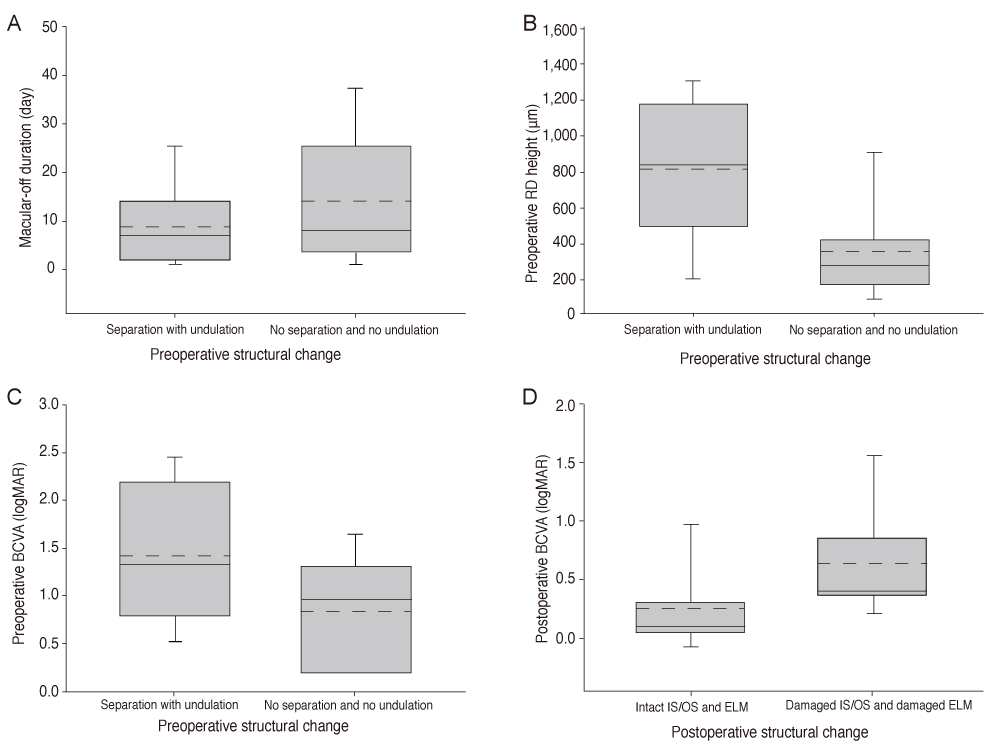
Fig. 2 (A) No significant difference in macula-off duration was evident between eyes with both intraretinal separation (IRS) and outer layer undulation (OLU) (p = 0.28). (B) Eyes with both IRS and OLU had a greater height of retinal detachment (RD) than did eyes lacking both IRS and OLU (p = 0.002). (C) Eyes with both IRS and OLU had lower best-corrected visual acuity (BCVA) values than eyes without either feature (p = 0.022). (D) Eyes with an intact junction between the photoreceptor inner and outer segments (IS/OS) and an intact external limiting membrane (ELM) showed better postoperative BCVA values than did eyes in which the IS/OS junction and the ELM were both damaged (p = 0.025). The solid lines indicate the medians and the long dashes the averages. logMAR = logarithm of the minimum angle of resolution.

Fig. 3 Eyes with both intraretinal separation (IRS) and outer layer undulation (OLU) exhibited lower integrity of both junction between the photoreceptor inner and outer segments and the external limiting membrane (p = 0.017). PHRc = photoreceptor complex including external limiting membrane and junction between the photoreceptor inner and outer segments.
Reference
-
1. Lecleire-Collet A, Muraine M, Menard JF, Brasseur G. Predictive visual outcome after macula-off retinal detachment surgery using optical coherence tomography. Retina. 2005. 25:44–53.2. Hassan TS, Sarrafizadeh R, Ruby AJ, et al. The effect of duration of macular detachment on results after the scleral buckle repair of primary, macula-off retinal detachments. Ophthalmology. 2002. 109:146–152.3. Ross WH, Stockl FA. Visual recovery after retinal detachment. Curr Opin Ophthalmol. 2000. 11:191–194.4. Lee SY, Joe SG, Kim JG, et al. Optical coherence tomography evaluation of detached macula from rhegmatogenous retinal detachment and central serous chorioretinopathy. Am J Ophthalmol. 2008. 145:1071–1076.5. Lecleire-Collet A, Muraine M, Menard JF, Brasseur G. Evaluation of macular changes before and after successful retinal detachment surgery using stratus-optical coherence tomography. Am J Ophthalmol. 2006. 142:176–179.6. Abouzeid H, Wolfensberger TJ. Macular recovery after retinal detachment. Acta Ophthalmol Scand. 2006. 84:597–605.7. Wakabayashi T, Oshima Y, Fujimoto H, et al. Foveal microstructure and visual acuity after retinal detachment repair: imaging analysis by Fourier-domain optical coherence tomography. Ophthalmology. 2009. 116:519–528.8. Schulze-Bonsel K, Feltgen N, Burau H, et al. Visual acuities "hand motion" and "counting fingers" can be quantified with the freiburg visual acuity test. Invest Ophthalmol Vis Sci. 2006. 47:1236–1240.9. Minihan M, Tanner V, Williamson TH. Primary rhegmatogenous retinal detachment: 20 years of change. Br J Ophthalmol. 2001. 85:546–548.10. McPherson AR, O'Malley RE, Butner RW, Beltangady SS. Visual acuity after surgery for retinal detachment with macular involvement. Ann Ophthalmol. 1982. 14:639–645.11. Yazici B, Gelisken O, Avci R, Yucel A. Prediction of visual outcome after retinal detachment surgery using the Lotmar visometer. Br J Ophthalmol. 2002. 86:278–281.12. Friberg TR, Eller AW. Prediction of visual recovery after scleral buckling of macula-off retinal detachments. Am J Ophthalmol. 1992. 114:715–722.13. Hagimura N, Suto K, Iida T, Kishi S. Optical coherence tomography of the neurosensory retina in rhegmatogenous retinal detachment. Am J Ophthalmol. 2000. 129:186–190.14. Kiernan DF, Mieler WF, Hariprasad SM. Spectral-domain optical coherence tomography: a comparison of modern high-resolution retinal imaging systems. Am J Ophthalmol. 2010. 149:18–31.15. Nakanishi H, Hangai M, Unoki N, et al. Spectral-domain optical coherence tomography imaging of the detached macula in rhegmatogenous retinal detachment. Retina. 2009. 29:232–242.16. Smith AJ, Telander DG, Zawadzki RJ, et al. High-resolution Fourier-domain optical coherence tomography and microperimetric findings after macula-off retinal detachment repair. Ophthalmology. 2008. 115:1923–1929.17. Arroyo JG, Yang L, Bula D, Chen DF. Photoreceptor apoptosis in human retinal detachment. Am J Ophthalmol. 2005. 139:605–610.18. Schocket LS, Witkin AJ, Fujimoto JG, et al. Ultrahigh-resolution optical coherence tomography in patients with decreased visual acuity after retinal detachment repair. Ophthalmology. 2006. 113:666–672.19. Shimoda Y, Sano M, Hashimoto H, et al. Restoration of photoreceptor outer segment after vitrectomy for retinal detachment. Am J Ophthalmol. 2010. 149:284–290.20. Doyle E, Herbert EN, Bunce C, et al. How effective is macula-off retinal detachment surgery. Might good outcome be predicted? Eye (Lond). 2007. 21:534–540.21. Laatikainen L, Harju H, Tolppanen EM. Post-operative outcome in rhegmatogenous retinal detachment. Acta Ophthalmol (Copenh). 1985. 63:647–655.22. Salicone A, Smiddy WE, Venkatraman A, Feuer W. Visual recovery after scleral buckling procedure for retinal detachment. Ophthalmology. 2006. 113:1734–1742.23. Tani P, Robertson DM, Langworthy A. Prognosis for central vision and anatomic reattachment in rhegmatogenous retinal detachment with macula detached. Am J Ophthalmol. 1981. 92:611–620.24. Machemer R. Experimental retinal detachment in the owl monkey. II. Histology of retina and pigment epithelium. Am J Ophthalmol. 1968. 66:396–410.25. Rossetti A, Doro D, Manfre A, Midena E. Long-term follow-up with optical coherence tomography and microperimetry in eyes with metamorphopsia after macula-off retinal detachment repair. Eye (Lond). 2010. 24:1808–1813.
- Full Text Links
-
- Actions
-
Cited
- CITED
-
- Close
- Share
-
- Similar articles
-
- The Usefulness of Optical Coherence Tomography in Macula-off Retinal Detachment
- Visual Prognosis and Foveal Reattachment After Reattachment Surgery in Macula-off Retinal Detachment
- Foveal Retinal Detachment Diagnosed by Optical Coherence Tomography after Successful Retinal Detachment Surgery
- Foveal Reattachment After Scleral Buckling vs Vitrectomy for Macula-Off Retinal Detachment
- A Case of Retinal Contusion Combined Exudative Retinal Detachment Causing Delayed Visual Disturbance
