Stafne bone cavity and cone-beam computed tomography: a report of two cases
- Affiliations
-
- 1Department of Oral Medicine and Radiology, College of Dentistry, Qaseem Private College, Buraidah, Kingdom of Saudi Arabia. dr_eva1@yahoo.com
- KMID: 1797845
- DOI: http://doi.org/10.5125/jkaoms.2015.41.3.145
Abstract
- In 1942 Stafne reported 35 asymptomatic, radiolucent cavities that were unilaterally located in the posterior region of the mandible between the mandibular angle and the third molar, and below the mandibular canal. The term Stafne bone cavity (SBC) is now used for such asymptomatic lingual bone depressions of the lower jaw. Since then there have been many reports of SBCs but very fews tudies have used cone-beam computed tomography (CBCT) for their diagnosis. The aim of this paper is to describe the clinical and radiological characteristics of two cases of SBCs and the importance of limited CBCT in confirming the diagnosis.
MeSH Terms
Figure
-
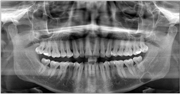
Fig. 1 Panoramic view shows the lesion.
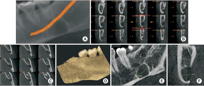
Fig. 2 Cone-beam computed tomography images. A. Panoramic view with mandibular nerve canal traced. B. Cross-section revealing the relationship of the lesion with the mandibular canal. C. Axial view revealing the relationship of the lesion with the mandibular canal. D. Three-dimensional reconstructed view from lingual aspect. E, F. Dimensions of the lesion.
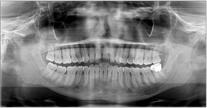
Fig. 3 Panoramic view shows the lesion.

Fig. 4 Cone-beam computed tomography images. A. Panoramic view with mandibular nerve canal traced. B. Cross-sections revealing the relationship of the lesion with the mandibular canal. C. Axial view revealing the relationship of the lesion with the mandibular canal. D. Three-dimensional reconstructed view from lingual aspect. E, F. Dimensions of the lesion.
Reference
-
1. Stafne EC. Bone cavities situated near the angle of the mandible. J Am Dent Assoc. 1942; 29:1969–1972.
Article2. Grellner TJ, Frost DE, Brannon RB. Lingual mandibular bone defect: report of three cases. J Oral Maxillofac Surg. 1990; 48:288–296. PMID: 2406401.
Article3. Boyle CA, Horner K, Coulthard P, Fleming GJ. Multiple Stafne bone cavities: a diagnostic dilemma. Dent Update. 2000; 27:494–497. PMID: 11218610.
Article4. Quesada-Gómez C, Valmaseda-Castellón E, Berini-Aytés L, Gay-Escoda C. Stafne bone cavity: a retrospective study of 11 cases. Med Oral Patol Oral Cir Bucal. 2006; 11:E277–E280. PMID: 16648768.5. Correll RW, Jensen JL, Rhyne RR. Lingual cortical mandibular defects: a radiographic incidence study. Oral Surg Oral Med Oral Pathol. 1980; 50:287–291. PMID: 6932004.6. Philipsen HP, Takata T, Reichart PA, Sato S, Suei Y. Lingual and buccal mandibular bone depressions: a review based on 583 cases from a world-wide literature survey, including 69 new cases from Japan. Dentomaxillofac Radiol. 2002; 31:281–290. PMID: 12203126.
Article7. Flores Campos PS, Oliveira JA, Dantas JA, de Melo DP, Pena N, Santos LA, et al. Stafne's defect with buccal cortical expansion: a case report. Int J Dent. 2010; DOI: 10.1155/2010/515931.
Article8. Sahin M, Gorgun S, Guven O. Stafne's bone cavity. Turkey Clinic Dental Sci J. 2005; 11:39–42.9. Dereci O, Duran S. Intraorally exposed anterior Stafne bone defect: a case report. Oral Surg Oral Med Oral Pathol Oral Radiol. 2012; 113:e1–e3. PMID: 22676987.
Article10. Turkoglu K, Orhan K. Stafne bone cavity in the anterior mandible. J Craniofac Surg. 2010; 21:1769–1775. PMID: 21119418.
Article11. Segev Y, Puterman M, Bodner L. Stafne bone cavity--magnetic resonance imaging. Med Oral Patol Oral Cir Bucal. 2006; 11:E345–E347. PMID: 16816811.12. Branstetter BF, Weissman JL, Kaplan SB. Imaging of a Stafne bone cavity: what MR adds and why a new name is needed. AJNR Am J Neuroradiol. 1999; 20:587–589. PMID: 10319966.13. Aguiar LB, Neves FS, Bastos LC, Crusoé-Rebello I, Ambrosano GM, Campos PS. Multiple stafne bone defects: a rare entity. ISRN Dent. 2011; DOI: 10.5402/2011/792145.
Article14. Layne EL, Morgan AF, Morton TH Jr. Anterior lingual mandibular bone concavity: report of case. J Oral Surg. 1981; 39:599–600. PMID: 6940969.15. Dorman M, Pierse D. Ectopic salivary gland tissue in the anterior mandible: a case report. Br Dent J. 2002; 193:571–572. PMID: 12481179.
Article16. de Courten A, Küffer R, Samson J, Lombardi T. Anterior lingual mandibular salivary gland defect (Stafne defect) presenting as a residual cyst. Oral Surg Oral Med Oral Pathol Oral Radiol Endod. 2002; 94:460–464. PMID: 12374920.
Article17. Barker GR. Xeroradiography in relation to a Stafne bone cavity. Br J Oral Maxillofac Surg. 1988; 26:32–35. PMID: 3422821.
Article18. Reuter I. An unusual case of Stafne bone cavity with extra-osseous course of the mandibular neurovascular bundle. Dentomaxillofac Radiol. 1998; 27:189–191. PMID: 9693534.
Article19. Ariji E, Fujiwara N, Tabata O, Nakayama E, Kanda S, Shiratsuchi Y, et al. Stafne's bone cavity. Classification based on outline and content determined by computed tomography. Oral Surg Oral Med Oral Pathol. 1993; 76:375–380. PMID: 8378054.
- Full Text Links
-
- Actions
-
Cited
- CITED
-
- Close
- Share
-
- Similar articles
-
- Management of root canal perforation by using cone-beam computed tomography
- Three-dimensional imaging modalities in endodontics
- Detection of maxillary second molar with two palatal roots using cone beam computed tomography: a case report
- Developmental salivary gland depression in the ascending mandibular ramus: A cone-beam computed tomography study
- Isolated tympanic plate fracture detected by cone-beam computed tomography: report of four cases with review of literature
