Odontogenic carcinosarcoma of the mandible: a case report and review
- Affiliations
-
- 1Department of Oral and Maxillofacial Surgery, Section of Dentistry, Inha University School of Medicine, Incheon, Korea. kik@inha.ac.kr
- 2Department of Pathology, Inha University School of Medicine, Incheon, Korea.
- KMID: 1797844
- DOI: http://doi.org/10.5125/jkaoms.2015.41.3.139
Abstract
- Odontogenic carcinosarcoma is an extremely rare malignant odontogenic tumor with only a few reported cases. It is characterized by a true mixed tumor showing malignant cytology of both epithelial and mesenchymal components. It has been assumed to arise from pre-existing lesions such as ameloblastoma, ameloblastic fibroma, and ameloblastic fibrosarcoma. To date, the reported cases have exhibited considerably aggressive clinical behavior. The case of an odontogenic carcinosarcoma in the mandible of a 61-year-old male is described herein. The tumor destroyed the cortex of the mandible and invaded the adjacent tissues. Treatment was performed by surgical resection and reconstruction. The purposes of this article are to introduce odontogenic carcinosarcoma through this case study, to distinguish it from related diseases and to discuss features of the tumor in the existing literature.
MeSH Terms
Figure
-
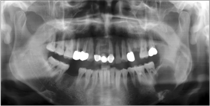
Fig. 1 Panoramic view shows radiolucent lesion of the left mandible.
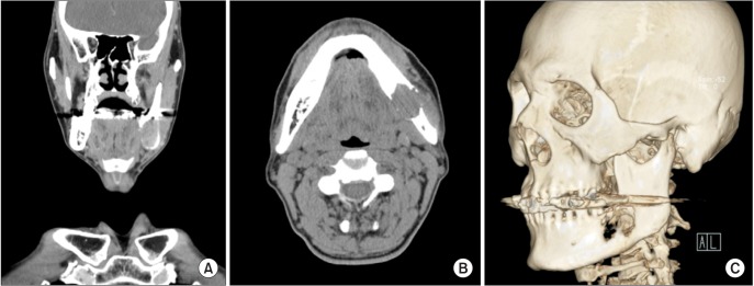
Fig. 2 Computed tomography shows an osteolytic mass with destruction of the inner cortex in the angle of the left mandible, involving the periapical area of the left 2nd molar tooth and left alveolar canal. A. Coronal view. B. Axial view. C. Three-dimensional image view.

Fig. 3 Magnetic resonance imaging shows 2.2×2.8×4.1 cm sized osteolyitc enhancing mass in the angle of the left mandible with invasion of the adjacent mandible bone marrow, surrounding tissue, and masseter muscle. A. Coronal view. B. Axial view.
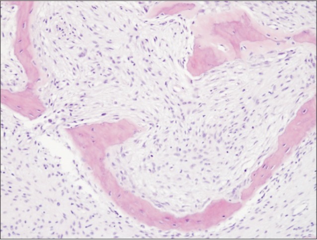
Fig. 4 Histopathologic image of the tibial lesion (H&E staining, ×200).

Fig. 5 Excised mass with 3.5×3.4×2.5 cm size which included tumor, mandible, and regional lymph node.
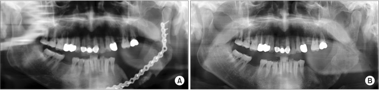
Fig. 6 A. Postoperative (1 month) panoramic view. B. Final postoperative (6 months) panoramic view after plate removal.
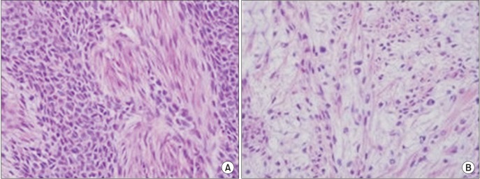
Fig. 7 A. Malignant epithelial component with pleomorphism, enlarged and atypical nuclei (H&E staining, ×400). B. Malignant mesenchymal component with hypercellularity, pleomorphism, enlarged and atypical nuclei (H&E staining, ×400).
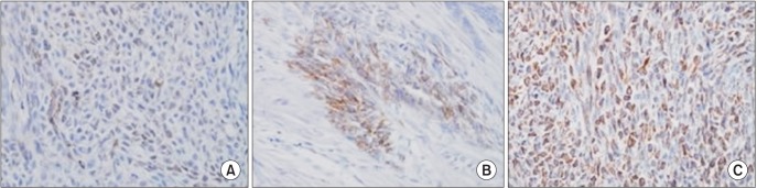
Fig. 8 Immunohistochemical stain images (×400). A. Malignant epithelial component. Intense membranous and cytoplasmic staining for cytokeratin. B. Positive staining for CAM 5.2 antigen supporting the diagnosis of malignancy. C. Malignant mesenchymal component showing positivity for vimentin.
Cited by 1 articles
-
Undifferentiated pleomorphic sarcoma of the mandible
Bernar Monteiro Benites, Wanessa Miranda-Silva, Felipe Paiva Fonseca, Claudia Regina Gomes Cardim Mendes de Oliveira, Eduardo Rodrigues Fregnani
J Korean Assoc Oral Maxillofac Surg. 2020;46(4):282-287. doi: 10.5125/jkaoms.2020.46.4.282.
Reference
-
1. Tanaka T, Ohkubo T, Fujitsuka H, Tatematsu N, Oka N, Kojima T, et al. Malignant mixed tumor (malignant ameloblastoma and fibrosarcoma) of the maxilla. Arch Pathol Lab Med. 1991; 115:84–87. PMID: 1987921.2. Slama A, Yacoubi T, Khochtali H, Bakir A. Mandibular odontogenic carcinosarcoma: a case report. Rev Stomatol Chir Maxillofac. 2002; 103:124–127. PMID: 11997741.3. Kunkel M, Ghalibafian M, Radner H, Reichert TE, Fischer B, Wagner W. Ameloblastic fibrosarcoma or odontogenic carcinosarcoma: a matter of classification? Oral Oncol. 2004; 40:444–449. PMID: 14969825.
Article4. DeLair D, Bejarano PA, Peleg M, El-Mofty SK. Ameloblastic carcinosarcoma of the mandible arising in ameloblastic fibroma: a case report and review of the literature. Oral Surg Oral Med Oral Pathol Oral Radiol Endod. 2007; 103:516–520. PMID: 17395065.
Article5. Chikosi R, Segall N, Augusto P, Freedman P. Odontogenic carcinosarcoma: case report and literature review. J Oral Maxillofac Surg. 2011; 69:1501–1507. PMID: 21195529.
Article6. Slootweg PJ. Malignant odontogenic tumors: an overview. Mund Kiefer Gesichtschir. 2002; 6:295–302. PMID: 12448230.
Article7. Chan WK, Li CP, Liu JM, Yin NT, Huang MH, Wu HP, et al. Mandibular odontogenic fibrosarcoma. Case report. Aust Dent J. 1997; 42:409–412. PMID: 9470285.
Article8. Sapp JP, Eversol LR, Wysocki GP. Contemporary oral and maxillofacial pathology. 2nd ed. New York: Elsevier Inc.;2004.9. Kramer IRH, Pindborg JJ, Shear M. Histological typing of odontogenic tumours. 2nd ed. New York: Springer;1992.
