Quantitative Computed Tomography of Pulmonary Emphysema and Ventricular Function in Chronic Obstructive Pulmonary Disease Patients with Pulmonary Hypertension
- Affiliations
-
- 1Institute of Biomedical Engineering, College of Medicine and College of Engineering, National Taiwan University, Taipei 100, Taiwan.
- 2Department of Medical Imaging, National Taiwan University Hospital and National Taiwan University College of Medicine, Taipei 100, Taiwan. ycc5566@ntu.edu.tw
- 3Department of Medical Imaging, National Taiwan University Hospital Yun Lin Branch, Yun-Lin 640, Taiwan.
- 4Department of Surgery, National Taiwan University Hospital and National Taiwan University College of Medicine, Taipei 100, Taiwan.
- KMID: 1794662
- DOI: http://doi.org/10.3348/kjr.2014.15.6.871
Abstract
OBJECTIVE
This study strived to evaluate the relationship between degree of pulmonary emphysema and cardiac ventricular function in chronic obstructive pulmonary disease (COPD) patients with pulmonary hypertension (PH) using electrocardiographic-gated multidetector computed tomography (CT).
MATERIALS AND METHODS
Lung transplantation candidates with the diagnosis of COPD and PH were chosen for the study population, and a total of 15 patients were included. The extent of emphysema is defined as the percentage of voxels below -910 Hounsfield units in the lung windows in whole lung CT without intravenous contrast. Heart function parameters were measured by electrocardiographic-gated CT angiography. Linear regression analysis was conducted to examine the associations between percent emphysema and heart function indicators.
RESULTS
Significant correlations were found between percent emphysema and right ventricular (RV) measurements, including RV end-diastolic volume (R2 = 0.340, p = 0.023), RV stroke volume (R2 = 0.406, p = 0.011), and RV cardiac output (R2 = 0.382, p = 0.014); the correlations between percent emphysema and left ventricular function indicators were not observed.
CONCLUSION
The study revealed that percent emphysema is correlated with RV dysfunction among COPD patients with PH. Based on our findings, percent emphysema can be considered for use as an indicator to predict the severity of right ventricular dysfunction among COPD patients.
Keyword
MeSH Terms
-
Adult
Aged
Electrocardiography
Female
Heart Ventricles/radiography
Humans
Hypertension, Pulmonary/complications/*diagnosis
Lung/radiography
Male
Middle Aged
Multidetector Computed Tomography
Pulmonary Disease, Chronic Obstructive/complications/*radiography
Pulmonary Emphysema/complications/*radiography
Regression Analysis
Ventricular Function/*physiology
Figure
-

Fig. 1 Snap shot of successful segmentation of each of cardiac chambers after semiautomatic processing and manual adjustment during different cardiac phases. Ventricular volume equals sum of all endocardial areas multiplied by slice thickness.
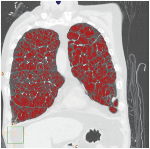
Fig. 2 Coronal reformatted lung window image in 57-year-old man with severe chronic obstructive pulmonary disease amenable to lung transplantation. Red areas indicate voxels below -910 Hounsfield units in lung parenchyma, which represent emphysematous change.
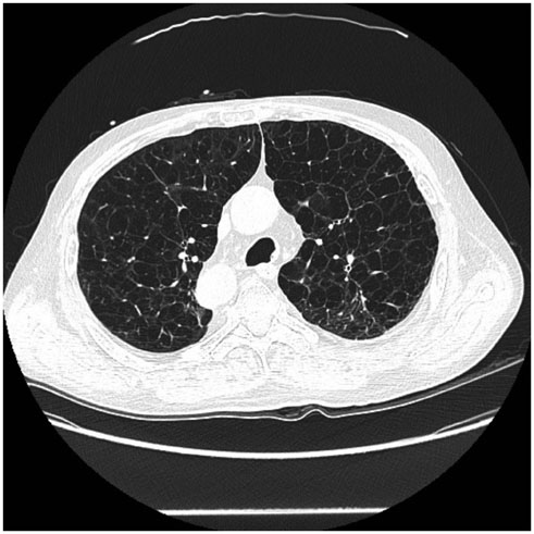
Fig. 3 Axial image of whole lung CT without intravenous contrast, in 60-year-old man with severe chronic obstructive pulmonary disease, demonstrates severe emphysema in lung window.
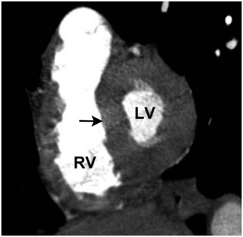
Fig. 4 Coronal short-axis image of contrast-enhanced CT in end-systole phase, in 67-year-old woman with severe chronic obstructive pulmonary disease, demonstrates straightening of interventricular septum (arrow) to left ventricle (LV) which indicates right ventricle (RV) dysfunction.
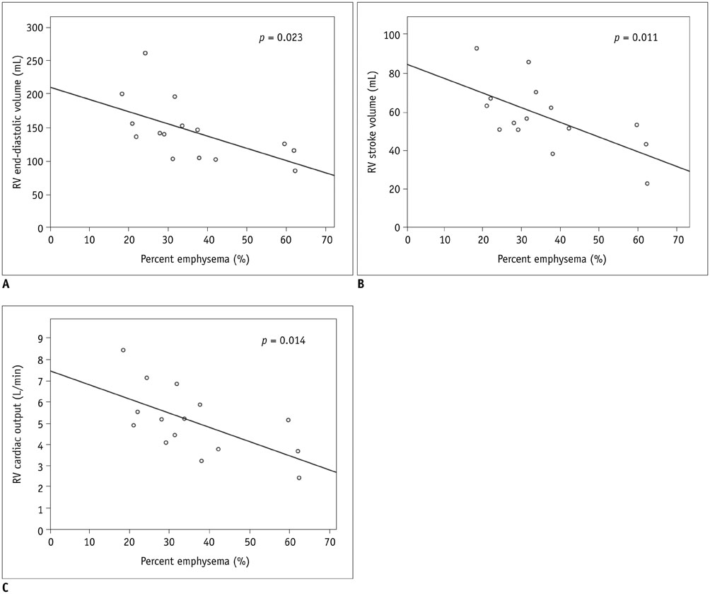
Fig. 5 Relationship of pulmonary percent emphysema and right ventricle parameters in chronic obstructive pulmonary disease patients with pulmonary hypertension. A. Right ventricular end-diastolic volume (R2 = 0.340, p = 0.023). B. Right ventricular stroke volume (R2 = 0.406, p = 0.011). C. Right ventricular cardiac output (R2 = 0.382, p = 0.014).
Cited by 1 articles
-
Quantitative Computed Tomography Assessment of Respiratory Muscles in Male Patients Diagnosed with Emphysema
Ji-Yeon Han, Ki-Nam Lee, Eun-Ju Kang, Jin Wook Baek
J Korean Soc Radiol. 2018;78(6):371-379. doi: 10.3348/jksr.2018.78.6.371.
Reference
-
1. MacNee W. Pathophysiology of cor pulmonale in chronic obstructive pulmonary disease. Part One. Am J Respir Crit Care Med. 1994; 150:833–852.2. Barberà JA, Peinado VI, Santos S. Pulmonary hypertension in chronic obstructive pulmonary disease. Eur Respir J. 2003; 21:892–905.3. Kasahara Y, Tuder RM, Cool CD, Lynch DA, Flores SC, Voelkel NF. Endothelial cell death and decreased expression of vascular endothelial growth factor and vascular endothelial growth factor receptor 2 in emphysema. Am J Respir Crit Care Med. 2001; 163(3 Pt 1):737–744.4. Jörgensen K, Müller MF, Nel J, Upton RN, Houltz E, Ricksten SE. Reduced intrathoracic blood volume and left and right ventricular dimensions in patients with severe emphysema: an MRI study. Chest. 2007; 131:1050–1057.5. Jörgensen K, Houltz E, Westfelt U, Nilsson F, Scherstén H, Ricksten SE. Effects of lung volume reduction surgery on left ventricular diastolic filling and dimensions in patients with severe emphysema. Chest. 2003; 124:1863–1870.6. Shin KE, Chung MJ, Jung MP, Choe BK, Lee KS. Quantitative computed tomographic indexes in diffuse interstitial lung disease: correlation with physiologic tests and computed tomography visual scores. J Comput Assist Tomogr. 2011; 35:266–271.7. Raman SV, Shah M, McCarthy B, Garcia A, Ferketich AK. Multi-detector row cardiac computed tomography accurately quantifies right and left ventricular size and function compared with cardiac magnetic resonance. Am Heart J. 2006; 151:736–744.8. Gao Y, Du X, Liang L, Cao L, Yang Q, Li K. Evaluation of right ventricular function by 64-row CT in patients with chronic obstructive pulmonary disease and cor pulmonale. Eur J Radiol. 2012; 81:345–353.9. Barr RG, Bluemke DA, Ahmed FS, Carr JJ, Enright PL, Hoffman EA, et al. Percent emphysema, airflow obstruction, and impaired left ventricular filling. N Engl J Med. 2010; 362:217–227.10. Grau M, Barr RG, Lima JA, Hoffman EA, Bluemke DA, Carr JJ, et al. Percent emphysema and right ventricular structure and function: the Multi-Ethnic Study of Atherosclerosis-Lung and Multi-Ethnic Study of Atherosclerosis-Right Ventricle Studies. Chest. 2013; 144:136–144.11. Sachdev A, Villarraga HR, Frantz RP, McGoon MD, Hsiao JF, Maalouf JF, et al. Right ventricular strain for prediction of survival in patients with pulmonary arterial hypertension. Chest. 2011; 139:1299–1309.12. Fisher MR, Criner GJ, Fishman AP, Hassoun PM, Minai OA, Scharf SM, et al. Estimating pulmonary artery pressures by echocardiography in patients with emphysema. Eur Respir J. 2007; 30:914–921.13. Orens JB, Estenne M, Arcasoy S, Conte JV, Corris P, Egan JJ, et al. International guidelines for the selection of lung transplant candidates: 2006 update--a consensus report from the Pulmonary Scientific Council of the International Society for Heart and Lung Transplantation. J Heart Lung Transplant. 2006; 25:745–755.14. Remy-Jardin M, Delhaye D, Teisseire A, Hossein-Foucher C, Duhamel A, Remy J. MDCT of right ventricular function: impact of methodologic approach in estimation of right ventricular ejection fraction, part 2. AJR Am J Roentgenol. 2006; 187:1605–1609.15. Coxson HO, Mayo JR, Behzad H, Moore BJ, Verburgt LM, Staples CA, et al. Measurement of lung expansion with computed tomography and comparison with quantitative histology. J Appl Physiol (1985). 1995; 79:1525–1530.16. Boussuges A, Pinet C, Molenat F, Burnet H, Ambrosi P, Badier M, et al. Left atrial and ventricular filling in chronic obstructive pulmonary disease. An echocardiographic and Doppler study. Am J Respir Crit Care Med. 2000; 162(2 Pt 1):670–675.17. Hur J, Kim TH, Kim SJ, Ryu YH, Kim HJ. Assessment of the right ventricular function and mass using cardiac multi-detector computed tomography in patients with chronic obstructive pulmonary disease. Korean J Radiol. 2007; 8:15–21.18. Watz H, Waschki B, Meyer T, Kretschmar G, Kirsten A, Claussen M, et al. Decreasing cardiac chamber sizes and associated heart dysfunction in COPD: role of hyperinflation. Chest. 2010; 138:32–38.19. Berger HJ, Matthay RA. Noninvasive radiographic assessment of cardiovascular function in acute and chronic respiratory failure. Am J Cardiol. 1981; 47:950–962.20. Hara AK, Paden RG, Silva AC, Kujak JL, Lawder HJ, Pavlicek W. Iterative reconstruction technique for reducing body radiation dose at CT: feasibility study. AJR Am J Roentgenol. 2009; 193:764–771.
- Full Text Links
-
- Actions
-
Cited
- CITED
-
- Close
- Share
-
- Similar articles
-
- Pulmonary Emphysema: Visual Interpretation and Quantitative Analysis
- Clinical use of chest CT in chronic obstructive pulmonary diseases
- Echocardiographic Evaluation of Right Ventricular Diastolic Function in Patients with Chronic Obstructive Pulmonary Disease
- Pathophysiology of Chronic Obstructive Pulmonary Disease
- Pathophysiology of Chronic Obstructive Pulmonary Disease
