Silicosis Caused by Chronic Inhalation of Snail Shell Powder
- Affiliations
-
- 1Department of Internal Medicine, Chung-Ang University College of Medicine, Seoul, Korea. jykimmd@cau.ac.kr
- KMID: 1792986
- DOI: http://doi.org/10.3346/jkms.2012.27.1.93
Abstract
- A 70-yr-old woman visited our hospital for shortness of breath. Chest CT showed ground glass opacity and traction bronchiectasis at right middle, lower lobe and left lingular division. Video-assisted thoracic surgical biopsy at right lower lobe and pathologic examination revealed mixed dust pneumoconiosis. Polarized optical microscopy showed lung lesions were consisted of silica and carbon materials. She was a housewife and never been exposed to silica dusts occupationally. She has taken freshwater snails as a health-promoting food for 40 yr and ground shell powder was piled up on her backyard where she spent day-time. Energy dispersive X-ray spectroscopy of snail shell and scanning electron microscopy with energy dispersive x-ray spectroscopy of lung lesion revealed that silica occupies important portion. Herein, we report the first known case of silicosis due to chronic inhalation of shell powder of freshwater snail.
Keyword
MeSH Terms
Figure
-
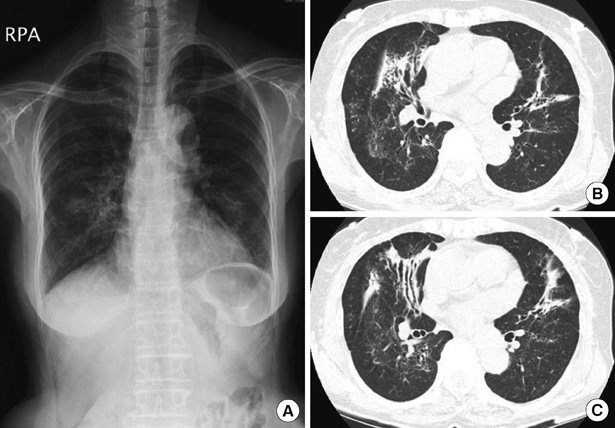
Fig. 1 Radiologic findings of the patent. (A) Chest PA demonstrates peribronchial infiltrations at middle and lower lung zones. (B, C) Chest CT shows ground glass opacity and traction bronchiectasis at right middle & lower lobe and left upper lobe lingular division.
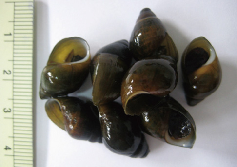
Fig. 2 Korean freshwater snails, Semisulcospira libertina, which the patient has taken for 40 yr (The reference of a scale bar is cm).

Fig. 3 Pathologic examination of lung sample taken by video-assisted thoracic surgery. (A) The multiple fibrotic nodules are distributed in perivascular and peribronchiolar interstitia on low power view (H&E, original × 40). (B) The nodules are composed of mixtures of collagenized fibroblasts and dust-laden macrophages (H&E, original × 400). (C) Partial polarization of mixed dust fibrotic nodules demonstrates brightly bi-refringent silicate particles (polarized optical microscopy, original × 400).
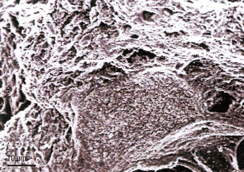
Fig. 4 The scattered platy and polymorphous silicate particles are observed by the scanning electron microscopic examination.
Reference
-
1. Scarisbrick D. Silicosis and coal workers' pneumoconiosis. Practitioner. 2002. 246:117–119.2. Honma K, Abraham JL, Chiyotani K, De Vuyst P, Dumortier P, Gibbs AR, Green FH, Hosoda Y, Iwai K, Williams WJ, Kohyama N, Ostiguy G, Roggli VL, Shida H, Taguchi O, Vallyathan V. Proposed criteria for mixed-dust pneumoconiosis: definition, descriptions, and guidelines for pathologic diagnosis and clinical correlation. Hum Pathol. 2004. 35:1515–1523.3. Arakawa H, Johkoh T, Honma K, Saito Y, Fukushima Y, Shida H, Suganuma N. Chronic interstitial pneumonia in silicosis and mix-dust pneumoconiosis: its prevalence and comparison of CT findings with idiopathic pulmonary fibrosis. Chest. 2007. 131:1870–1876.4. 't Mannetje A, Steenland K, Attfield M, Boffetta P, Checkoway H, DeKlerk N, Koskela RS. Exposure-response analysis and risk assessment for silica and silicosis mortality in a pooled analysis of six cohorts. Occup Environ Med. 2002. 59:723–728.5. Castranova V, Vallyathan V. Silicosis and coal workers' pneumoconiosis. Environ Health Perspect. 2000. 108:675–678.6. Schenker M. Exposures and health effects from inorganic agricultural dusts. Environ Health Perspect. 2000. 108:661–664.7. Sherwin RP, Barman ML, Abraham JL. Silicate pneumoconiosis of farm workers. Lab Invest. 1979. 40:576–582.
