Epithelial-Mesenchymal Transitions of Bile Duct Epithelial Cells in Primary Hepatolithiasis
- Affiliations
-
- 1Hepatobiliary Surgery Institute, Southwest Hospital, Third Military Medical University, Chongqing, China. sgwang90@yahoo.com
- 2Affiliated Hospital of Zunyi Medical College, Zunyi, China.
- 3Department of General Surgery, The 324th Hospital of PLA, Chongqing, China.
- KMID: 1792958
- DOI: http://doi.org/10.3346/jkms.2010.25.7.1066
Abstract
- The purpose of this study was to explore the role of epithelial-mesenchymal transition in the pathogenesis of hepatolithiasis. Thirty-one patients with primary hepatolithiasis were enrolled in this study. Expressions of E-cadherin, alpha-catenin, alpha-SMA, vimentin, S100A4, TGF-beta1 and P-smad2/3 in hepatolithiasis bile duct epithelial cells were examined by immunohistochemistry staining. The results showed that the expressions of the epithelial markers E-cadherin and alpha-catenin were frequently lost in hepatolithiasis (32.3% and 25.9% of cases, respectively), while the mesenchymal markers vimentin, alpha-SMA and S100A4 were found to be present in hepatolithiasis (35.5%, 29.0%, and 32.3% of cases, respectively). The increased mesenchymal marker expression was correlated with decreased epithelial marker expression. The expressions of TGF-beta1 and P-smad2/3 in hepatolithiasis were correlated with the expression of S100A4. These data indicate that TGF-beta1-mediated epithelial-mesenchymal transition might be involved in the formation of hepatolithiasis.
Keyword
MeSH Terms
Figure
-
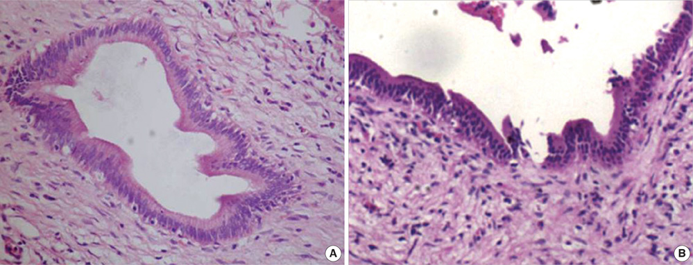
Fig. 1 Histopathological assessment of HL. Histopathological assessment (hematoxylin-eosin staining) of samples from (A) control group and (B) primary hepatolithiasis, abnormalities in which include infiltrating inflammatory cells in portal areas, fibrous tissue hyperplasia, biliary dilatation, and necrosis. Magnification ×200.
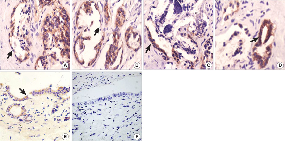
Fig. 2 Immunohistochemical assessment of epithelial and mesenchymal markers. Expressions of the epithelial markers (A) E-cadherin and (B) α-catenin in primary hepatolithiasis. Some BECs lost expression of epithelial markers (arrows). Expressions of mesenchymal markers (C) Vimentin, (D) α-SMA and (E) S100A4 in the liver bile ducts in hepatolithiasis. Note the brown staining of the markers lining the plasma membrane and in the cytoplasm (arrows). In normal liver tissue, S100A4 is neganative (F). Magnification ×400.
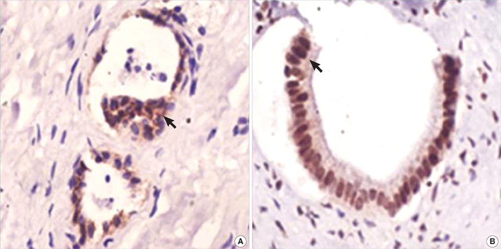
Fig. 3 Immunohistochemical assessment of TGF-β1 and P-smad2/3. (A) TGF-β1 expressed in the cytoplasm and on the plasma membrane of bile duct epithelial cells in hepatolithiasis, and (B) P-smad2/3 accumulated in the nucleus of bile duct epithelial cells in hepatolithiasis. Magnification ×400.
Reference
-
1. Shoda J, Tanaka N, Osuga T. Hepatolithiasis-epidemiology and pathogenesis update. Front Biosci. 2003. 8.
Article2. Kubo S, Kinoshita H, Hirohashi K, Hamba H. Hepatolithiasis associated with cholangiocarcinoma. World J Surg. 1995. 19:637–641.
Article3. Nakai A, Imano M, Takeyama Y, Shiozaki H, Ohyanagi H. An immunohistochemical study of osteopontin in hepatolithiasis. J Hepatobil Pancreat Surg. 2008. 15:615–621.
Article4. Thiery JP. Epithelial-mesenchymal transitions in tumour progression. Nat Rev Cancer. 2002. 2:442–454.
Article5. Vongwiwatana A, Tasanarong A, Rayner DC, Melk A, Halloran PF. Epithelial to mesenchymal transition during late deterioration of human kidney transplants: the role of tubular cells in fibrogenesis. Am J Transplant. 2005. 5:1367–1374.
Article6. Robertson H, Kirby JA, Yip WW, Jones DE, Burt AD. Biliary epithelial-mesenchymal transition in posttransplantation recurrence of primary biliary cirrhosis. Hepatology. 2007. 45:977–981.
Article7. Schiffer M, von Gersdorff G, Bitzer M, Susztak K, Bottinger EP. Smad proteins and transforming growth factor-beta signaling. Kidney Int. 2000. 77:Suppl. S45–S52.8. ten Dijke P, Hill CS. New insights into TGF-beta-Smad signalling. Trends Biochem Sci. 2004. 29:265–273.9. Rygiel KA, Robertson H, Marshall HL, Pekalski M, Zhao L, Booth TA, Jones DE, Burt AD, Kirby JA. Epithelial-mesenchymal transition contributes to portal tract fibrogenesis during human chronic liver disease. Lab Invest. 2008. 88:112–123.
Article10. Han DB, Dong JH, Guo GJ. Evaluation of our clinicopathologic stage for 1259 patients with intrahepatic stones. Acta Academiae Medicinae Militaris Tertiae. 2006. 28:1337–1338.11. Asayama Y, Taguchi Ki K, Aishima Si S, Nishi H, Masuda K, Tsuneyoshi M. The mode of tumour progression in combined hepatocellular carcinoma and cholangiocarcinoma: an immunohistochemical analysis of E-cadherin, alpha-catenin and beta-catenin. Liver. 2002. 22:43–50.
Article12. Nakajima S, Doi R, Toyoda E, Tsuji S, Wada M, Koizumi M, Tulachan SS, Ito D, Kami K, Mori T, Kawaguchi Y, Fujimoto K, Hosotani R, Imamura M. N-cadherin expression and epithelial-mesenchymal transition in pancreatic carcinoma. Clin Cancer Res. 2004. 10:4125–4133.
Article13. Kang Y, Massague J. Epithelial-mesenchymal transitions: twist in development and metastasis. Cell. 2004. 118:277–279.14. Lee TY, Chen YL, Chang HC, Chan CP, Kuo SJ. Outcomes of hepatectomy for hepatolithiasis. World J Surg. 2007. 31:479–482.
Article15. Inayoshi J, Ichida T, Sugitani S, Tsuboi Y, Genda T, Honma N, Asakura H. Gross appearance of hepatocellular carcinoma reflects E-cadherin expression and risk of early recurrence after surgical treatment. J Gastroenterol Hepatol. 2003. 18:673–677.
Article16. Yang J, Mani SA, Donaher JL, Ramaswamy S, Itzykson RA, Come C, Savagner P, Gitelman I, Richardson A, Weinberg RA. Twist, a master regulator of morphogenesis, plays an essential role in tumor metastasis. Cell. 2004. 117:927–939.
Article17. Zeisberg M, Bonner G, Maeshima Y, Colorado P, Muller GA, Strutz F, Kalluri R. Renal fibrosis: collagen composition and assembly regulates epithelial-mesenchymal transdifferentiation. Am J Pathol. 2001. 159:1313–1321.18. Choi HS, Savard CE, Choi JW, Kuver R, Lee SP. Paclitaxel interrupts TGF-beta1 signaling between gallbladder epithelial cells and myofibroblasts. J Surg Res. 2007. 141:183–191.
- Full Text Links
-
- Actions
-
Cited
- CITED
-
- Close
- Share
-
- Similar articles
-
- The Observation of Histologic Changes of Major Intrahepatic Bile Duct Epithelium in the Resected Liver Tissue with Hepatolithiasis
- Expression of Proliferating Cell Nuclear Antigen (PCNA) of Major Intrahepatic Bile Duct Epithelium in Resected Liver Tissue with Hepatolithiasis and Hepatolithiasis Associated with Cholangiocarcinoma
- The Crucial Role of Cholangiocytes in Cholangiopathies
- The Surgical Treatments for the Hepatolithiasis
- Histochemistry of Six Lectins in the Tissues of the Flat Fish Paralichthys olivaceus
