Esophageal Pyogenic Granuloma: Endosonographic Findings and Endoscopic Treatments
- Affiliations
-
- 1Department of Internal Medicine, Pusan National University School of Medicine and Biomedical Research Institute, Pusan National University Hospital, Busan, Korea. doc0224@pusan.ac.kr
- KMID: 1792645
- DOI: http://doi.org/10.5946/ce.2013.46.1.81
Abstract
- Pyogenic granuloma is a benign inflammatory vascular lesion, mainly found in the skin and oral mucosa. A few cases of pyogenic granuloma in the gastrointestinal tract have been reported, and the esophagus was the main site in these cases. These patients were diagnosed with pyogenic granuloma after they underwent upper endoscopy and biopsy. Endoscopic resection is a favorable treatment option for esophageal pyogenic granuloma. Recently, we observed characteristic endosonographic findings in two cases with esophageal pyogenic granuloma, which were then treated successfully by endoscopic resection.
Keyword
MeSH Terms
Figure
-
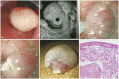
Fig. 1 (A) Endoscopy findings. A 1-cm polypoid lesion covered by exudates is observed just above the esophagogastric junction. (B) Endosonographic findings. A homogeneously hyperechoic lesion is seen in the submucosa. (C) Saline solution is injected to lift the lesion. (D) Complete removal of the lesion using a snare. (E) Resected specimen. (F) Histological findings. The resected polyp is composed of edematous granulated tissue containing numerous capillaries (H&E stain, ×200; Box, H&E stain, ×40).

Fig. 2 (A) Endoscopy findings. A 0.5-cm pinkish polypoid lesion is seen 30 cm from the incisor teeth. (B) Endosonographic findings. A homogeneously hyperechoic lesion is seen in the lamina propria. (C) Band ligation was performed using a ligation device and then snare resection was done. (D) Complete resection of the lesion. (E) Resected specimen. (F) Histological findings. The resected polyp is composed of abundant capillaries that are lined with endothelial cells (H&E stain, ×200; Box, H&E stain, ×40).
Reference
-
1. Poncet A, Dor L. Botryomycose humaine. Rev Chir. 1897; 18:996–1003.2. Park SY, Park CH, Lee WS, Kim HS, Choi SK, Rew JS. Pyogenic granuloma of the duodenum treated successfully by endoscopic mucosal resection. Gut Liver. 2009; 3:48–51. PMID: 20479901.
Article3. Moffatt DC, Warwryko P, Singh H. Pyogenic granuloma: an unusual cause of massive gastrointestinal bleeding from the small bowel. Can J Gastroenterol. 2009; 23:261–264. PMID: 19373418.
Article4. Kusakabe A, Kato H, Hayashi K, et al. Pyogenic granuloma of the stomach successfully treated by endoscopic resection after transarterial embolization of the feeding artery. J Gastroenterol. 2005; 40:530–535. PMID: 15942720.
Article5. Cho HS, Jung ES, Lee YJ, et al. A case of esophageal pyogenic granuloma. Korean J Gastrointest Endosc. 2009; 38:210–213.6. Chung IS, Kim SW, Choi MG, et al. A case of pyogenic granuloma in the terminal ileum treated by endoscopic snare polypectomy. Korean J Gastrointest Endosc. 2002; 24:176–180.7. Kerr DA. Granuloma pyogenicum. Oral Surg Oral Med Oral Pathol. 1951; 4:158–176. PMID: 14807485.
Article8. Bhaskar SN, Jacoway JR. Pyogenic granuloma: clinical features, incidence, histology, and result of treatment: report of 242 cases. J Oral Surg. 1966; 24:391–398. PMID: 5220911.
