Korean J Ophthalmol.
2014 Apr;28(2):122-129. 10.3341/kjo.2014.28.2.122.
The Correlation of Routine Tear Function Tests and Conjunctival Impression Cytology in Dry Eye Syndrome
- Affiliations
-
- 1Department of Pathology, Santosh Hospital, Santosh Medical College, Ghaziabad, India.
- 2Department of Ophthalmology, Santosh Hospital, Santosh Medical College, Ghaziabad, India. brahul_2371@yahoo.co.in
- 3Department of Microbiology, Narayan Medical College Hospital, Sasaram, India.
- KMID: 1792105
- DOI: http://doi.org/10.3341/kjo.2014.28.2.122
Abstract
- PURPOSE
To establish the strength of the association between routine tear function tests and conjunctival impression cytology (CIC) and to determine whether they simulate the morphological and cytological changes that occur on the ocular surface in dry eye. What are the sensitivity, specificity and positive predictive values of these tests when CIC is considered the gold standard?
METHODS
The tear film profile included tear film break up time (TBUT), Schirmer's-1, Rose Bengal scores (RBS), and impression cytology. CIC samples were obtained from the inferior bulbar conjunctiva and stained with periodic acid-Schiff and counter stained with hematoxylin and eosin.
RESULTS
The mean Schirmer's value was 11.66 +/- 5.90 in patients and 17.17 +/- 2.97 in controls (p < 0.001). The mean TBUT in participants was 8.88 +/- 3.54 and 13.53 +/- 2.12 in controls (p < 0.001). Patients had a mean goblet cell density (GCD) of 490 +/- 213, while the value for controls was 1,462 +/- 661 (p < 0.001). Abnormal CIC was observed in 46.7% cases of dry eye and in 32.8% of controls. The correlation coefficient (L) for Schirmer's was 0.2 and 0.24 for participants and controls, respectively, while TBUT values were 0.26 and 0.38, RBS were 0.5 and 0.5, and GCD was 0.8 and 0.6 in cases and controls, respectively.
CONCLUSIONS
GCD, RBS, and TBUT were better predictors of morphological and cytological changes in the conjunctiva than Schirmer's in dry eye syndrome. The sensitivity of tear function tests in diagnosing dry eye was TBUT > Schirmer's > RBS, and the specificity was Schirmer's > TBUT > RBS in decreasing order when CIC was considered the gold standard.
Keyword
MeSH Terms
-
Adolescent
Adult
Case-Control Studies
Child
Conjunctiva/pathology
Diagnostic Techniques, Ophthalmological/*standards
Dry Eye Syndromes/*diagnosis/*pathology
Eosine Yellowish-(YS)/diagnostic use
Female
Goblet Cells/pathology
Hematoxylin/diagnostic use
Humans
Male
Middle Aged
Ophthalmology/*standards
Periodic Acid-Schiff Reaction/standards
Predictive Value of Tests
Rose Bengal/diagnostic use
Sensitivity and Specificity
*Tears
Young Adult
Eosine Yellowish-(YS)
Hematoxylin
Rose Bengal
Figure
-
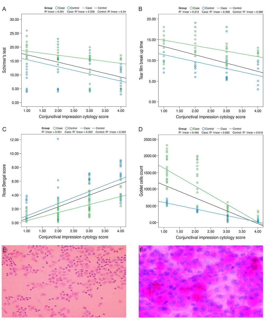
Fig. 1 (A) Scatter diagram showing correlation between Schirmer's test and conjunctival impression cytology. (B) Scatter diagram showing correlation between tear film break up time and conjunctival impression cytology. (C) Scatter diagram showing correlation between Rose Bengal score and conjunctival impression cytology. (D) Scatter diagram showing correlation between goblet cell density and conjunctival impression cytology. (E) Periodic acid-Schiff stained impression cytology specimen with mild dry eye and a reduced goblet cell count. (F) Periodic acid-Schiff stained image with squamous metaplasia, inflammatory infiltration and a decrease in goblet cells.
Reference
-
1. The definition and classification of dry eye disease: report of the Definition and Classification Subcommittee of the International Dry Eye WorkShop (2007). Ocul Surf. 2007; 5:75–92.2. Nichols KK, Mitchell GL, Zadnik K. The repeatability of clinical measurements of dry eye. Cornea. 2004; 23:272–285.3. Patel S, Murray D, McKenzie A, et al. Effects of fluorescein on tear breakup time and on tear thinning time. Am J Optom Physiol Opt. 1985; 62:188–190.4. Cho P, Brown B, Lau C. Effect of fluorescein on the tear stability of Hong Kong-Chinese. Optom Vis Sci. 1996; 73:1–7.5. Argueso P, Tisdale A, Spurr-Michaud S, et al. Mucin characteristics of human corneal-limbal epithelial cells that exclude the rose bengal anionic dye. Invest Ophthalmol Vis Sci. 2006; 47:113–119.6. Dart J. Impression cytology of the ocular surface: research tool or routine clinical investigation? Br J Ophthalmol. 1997; 81:930.7. Egbert PR, Lauber S, Maurice DM. A simple conjunctival biopsy. Am J Ophthalmol. 1977; 84:798–801.8. Natadisastra G, Wittpenn JR, West KP Jr, et al. Impression cytology for detection of vitamin A deficiency. Arch Ophthalmol. 1987; 105:1224–1228.9. Puangsricharern V, Tseng SC. Cytologic evidence of corneal diseases with limbal stem cell deficiency. Ophthalmology. 1995; 102:1476–1485.10. Lee GA, Hirst LW. Ocular surface squamous neoplasia. Surv Ophthalmol. 1995; 39:429–450.11. Bhargava R, Kumar P, Kumar M, et al. A randomized controlled trial of omega-3 fatty acids in dry eye syndrome. Int J Ophthalmol. 2013; 6:811–816.12. Lemp MA. Report of the National Eye Institute/Industry workshop on Clinical Trials in Dry Eyes. CLAO J. 1995; 21:221–232.13. Van Bijsterveld OP. Diagnostic tests in the Sicca syndrome. Arch Ophthalmol. 1969; 82:10–14.14. Nelson JD. Impression cytology. Cornea. 1988; 7:71–81.15. Foulks GN. Challenges and pitfalls in clinical trials of treatments for dry eye. Ocul Surf. 2003; 1:20–30.16. Nichols KK, Nichols JJ, Mitchell GL. The lack of association between signs and symptoms in patients with dry eye disease. Cornea. 2004; 23:762–770.17. Adatia FA, Michaeli-Cohen A, Naor J, et al. Correlation between corneal sensitivity, subjective dry eye symptoms and corneal staining in Sjogren's syndrome. Can J Ophthalmol. 2004; 39:767–771.18. Bjerrum KB. Test and symptoms in keratoconjunctivitis sicca and their correlation. Acta Ophthalmol Scand. 1996; 74:436–441.19. Murube J, Rivas L. Impression cytology on conjunctiva and cornea in dry eye patients establishes a correlation between squamous metaplasia and dry eye clinical severity. Eur J Ophthalmol. 2003; 13:115–127.20. Reddy M, Reddy PR, Reddy SC. Conjunctival impression cytology in dry eye states. Indian J Ophthalmol. 1991; 39:22–24.21. Bandyopadhyay R, Nag D, Mondal SK, et al. Ocular surface disorder in pterygium: role of conjunctival impression cytology. Indian J Pathol Microbiol. 2010; 53:692–695.22. Sood S, Shukla R, Nada M, et al. Comparison of tear film profile, conjunctival impression cytology, and conjunctival biopsy in patients with dry eye. Asian J Ophthalmol. 2006; 8:24–27.23. Tseng SC. Staging of conjunctival squamous metaplasia by impression cytology. Ophthalmology. 1985; 92:728–733.24. Paschides CA, Petroutsos G, Psilas K. Correlation of conjunctival impression cytology results with lacrimal function and age. Acta Ophthalmol (Copenh). 1991; 69:422–425.25. Yaylali V, Ozyurt C. Comparison of tear function tests and impression cytology with the ocular findings in acne rosacea. Eur J Ophthalmol. 2002; 12:11–17.26. Doughty MJ. Goblet cells of the normal human bulbar conjunctiva and their assessment by impression cytology sampling. Ocul Surf. 2012; 10:149–169.27. Doughty MJ. Sampling area selection for the assessment of goblet cell density from conjunctival impression cytology specimens. Eye Contact Lens. 2012; 38:122–129.28. Gupta Y, Gupta M, Maheshwari R, et al. Xerosis meter-an electro-physiological device for quick screening of dry eyes. Nepal J Ophthalmol. 2009; 1:123–128.29. Rahman A, Yahya K, Ahmed T, et al. Validity of symptoms as a screening tool for dry eye. Pak J Ophthalmol. 2007; 23:198–203.
- Full Text Links
-
- Actions
-
Cited
- CITED
-
- Close
- Share
-
- Similar articles
-
- Clinical Usefulness of Conjunctival Brush Cytology in the Diagnosis of Dry Eye Syndrome
- Therapeutic Effect of Topical Testosterone Gel in Patients with Dry Eye Syndrome
- Changes of Tear Parameters after Using Cyclosporine A in Dry Eye with Thyroid Ophthalmopahty
- Tear Film and Ocular Surface Changes in Chronic Renal Failure Patients Undergoing Hemodialysis
- Normal Conjunctival Goblet Cell Density in Korean Measured by Impression Cytology
