Retinal Damage in Chloroquine Maculopathy, Revealed by High Resolution Imaging: A Case Report Utilizing Adaptive Optics Scanning Laser Ophthalmoscopy
- Affiliations
-
- 1Department of Ophthalmology, Kangdong Sacred Heart Hospital, Hallym University Medical Center, Seoul, Korea. sungpyo@hanafos.com
- 2Department of Ophthalmology, Columbia University Medical Center, New York, NY, USA.
- KMID: 1792101
- DOI: http://doi.org/10.3341/kjo.2014.28.1.100
Abstract
- A 53-year-old Asian woman was treated with hydroxychloroquine and chloroquine for lupus erythematosus. Within a few years, she noticed circle-shaped shadows in her central vision. Upon examination, the patient's visual acuity was 20 / 25 in both eyes. Humphrey visual field (HVF) testing revealed a central visual defect, and fundoscopy showed a ring-shaped area of parafoveal retinal pigment epithelium depigmentation. Fundus autofluorescence imaging showed a hypofluorescent lesion consistent with bull's eye retinopathy. Adaptive optics scanning laser ophthalmoscope (AO-SLO) revealed patch cone mosaic lesions, in which cones were missing or lost. In addition, the remaining cones consisted of asymmetrical shapes and sizes that varied in brightness. Unlike previous studies employing deformable mirrors for wavefront aberration correction, our AO-SLO approach utilized dual liquid crystal on silicon spatial light modulators. Thus, by using AO-SLO, we were able to create a photographic montage consisting of high quality images. Disrupted cone AO-SLO images were matched with visual field test results and functional deficits were associated with a precise location on the montage, which allowed correlation of histological findings with functional changes determined by HVF. We also investigated whether adaptive optics imaging was more sensitive to anatomical changes compared with spectral-domain optical coherence tomography.
Keyword
MeSH Terms
Figure
-

Fig. 1 Photographs of the right (A) and left (B) eyes. Fundus autofluorescent (FAF) images of the right (C) and left (D) eyes are also shown. The FAF image shows a hypofluorescent lesion in the foveal and perifoveal areas consistent with bull's eye retinopathy. A prominent hypofluorescent lesion is visible in the left eye, indicating a marked atrophy of the retinal pigment epithelium layer. The bull's eye pattern of depigmentation is also evident on fundus photography and fundus autofluorescent images.
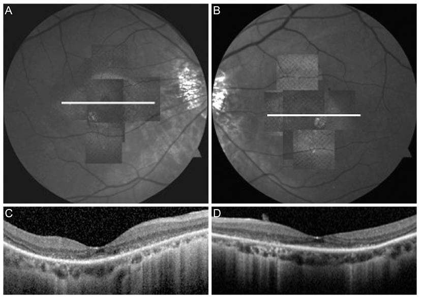
Fig. 2 Adaptive optics scanning laser ophthalmoscope (AO-SLO) montage (A,B) and spectral-domain optical coherence tomography (SD-OCT) (C,D) images of both eyes. The vertical SD-OCT images from both the right (A) and left (B) eyes show loss of photoreceptor inner segment/outer segment junctions (moth eaten appearance).
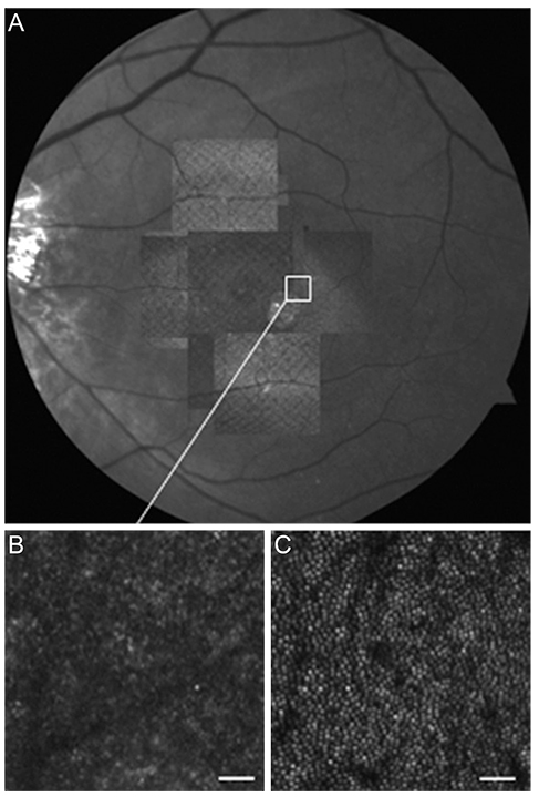
Fig. 3 Adaptive optics scanning laser ophthalmoscope (AO-SLO) montage from the left eye (A) matched with the corresponding red free image. Magnified AO-SLO images (B,C) are also shown. (B) shows the area indicated by the white box on the montage. For comparison, (C) shows an age-matched normal retina in the same location. As shown in (B), disruptions in the cone mosaic, where cones were missing or lost, is apparent. These disruptions were not present in the normal subject. Additionally, in (B), cones appear to be asymmetrical in shape and size with variable brightness. Scale bar in (B) and (C) = 25 µm.
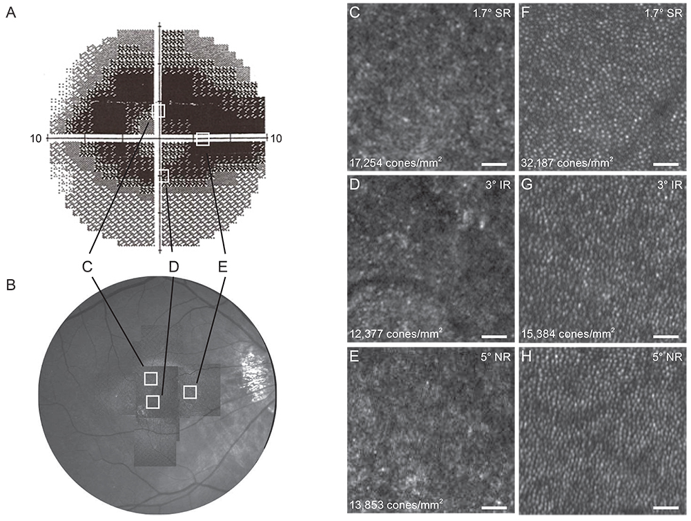
Fig. 4 Correlation of structural and functional defects. Humphrey visual field (A) revealed a significant central defect. The adaptive optics scanning laser ophthalmoscope (AO-SLO) montage from the right eye (B) was matched with the infrared image. Images (C), (D), and (E) are magnified AO-SLO images of the locations of visual field defects (white boxes). Images (F), (G), and (H) are images in the same location from an age-matched normal retina. The location and cone density for each figure is shown. The AO-SLO images (C), (D), and (E), show the cone mosaic disruption and dark patchy lesions where cones are missing or lost. Image D had the lowest cone density, and was lower than observed in a normal subject (F,G,H). Cones in images (C), (D), and (E) were asymmetric in shape and size and exhibited variable brightness. Scale bar in (C), (D), and (E) = 25 µm. SR = superior retina; IR = inferior retina; NR = nasal retina.
Reference
-
1. Rynes RI. Antimalarial drugs in the treatment of rheumatological diseases. Br J Rheumatol. 1997; 36:799–805.2. Mavrikakis M, Papazoglou S, Sfikakis PP, et al. Retinal toxicity in long term hydroxychloroquine treatment. Ann Rheum Dis. 1996; 55:187–189.3. Marmor MF, Carr RE, Easterbrook M, et al. Recommendations on screening for chloroquine and hydroxychloroquine retinopathy: a report by the American Academy of Ophthalmology. Ophthalmology. 2002; 109:1377–1382.4. Rosenthal AR, Kolb H, Bergsma D, et al. Chloroquine retinopathy in the rhesus monkey. Invest Ophthalmol Vis Sci. 1978; 17:1158–1175.5. Wetterholm DH, Winter FC. Histopathology of chloroquine retinal toxicity. Arch Ophthalmol. 1964; 71:82–87.6. Roorda A, Romero-Borja F, Donnelly W III, et al. Adaptive optics scanning laser ophthalmoscopy. Opt Express. 2002; 10:405–412.7. Park SP, Chung JK, Greenstein V, et al. A study of factors affecting the human cone photoreceptor density measured by adaptive optics scanning laser ophthalmoscope. Exp Eye Res. 2013; 108:1–9.8. Hirose F, Nozato K, Saito K, Numajiri Y. A compact adaptive optics scanning laser ophthalmoscope with high-efficiency wavefront correction using dual liquid crystal on silicon: spatial light modulator. Proc SPIE. 2011; 7885. doi: 10.1117/12.873671.9. Easterbrook M. Long-term course of antimalarial maculopathy after cessation of treatment. Can J Ophthalmol. 1992; 27:237–239.10. Schmidt B, Mueller-Limmroth W. Electroretinographic examinations following the application of chloroquine. Acta Ophthalmol Suppl. 1962; Suppl 70. 245–251.11. Henkind P, Carr RE, Siegel IM. Early chloroquine retinopathy: clinical and functional findings. Arch Ophthalmol. 1964; 71:157–165.12. Penrose PJ, Tzekov RT, Sutter EE, et al. Multifocal electroretinography evaluation for early detection of retinal dysfunction in patients taking hydroxychloroquine. Retina. 2003; 23:503–512.13. Park SP, Hong IH, Tsang SH, et al. Disruption of the human cone photoreceptor mosaic from a defect in NR2E3 transcription factor function in young adults. Graefes Arch Clin Exp Ophthalmol. 2013; 251:2299–2309.14. Park SP, Hong IH, Tsang SH, Chang S. Cellular imaging demonstrates genetic mosaicism in heterozygous carriers of an X-linked ciliopathy gene. Eur J Hum Genet. 2013; 21:1240–1248.15. Curcio CA, Sloan KR, Kalina RE, Hendrickson AE. Human photoreceptor topography. J Comp Neurol. 1990; 292:497–523.16. Stepien KE, Han DP, Schell J, et al. Spectral-domain optical coherence tomography and adaptive optics may detect hydroxychloroquine retinal toxicity before symptomatic vision loss. Trans Am Ophthalmol Soc. 2009; 107:28–33.
- Full Text Links
-
- Actions
-
Cited
- CITED
-
- Close
- Share
-
- Similar articles
-
- Principles of Adaptive Optics and Clinical Applications
- Whole Layer Photocoagulation on the Rabbit Retina with Indirect Diode Laser Ophthalmoscopy
- Comparison of Blue and Green Confocal Scanning Laser Ophthalmoscope Imaging to Detect Retinal Nerve Fiber Layer Defects
- The Optical Coherence Tomography Analysis of Hyperfluorescence by Scanning Laser Ophthalmoscope in Central Serous Chorioretinopathy
- Maculopathy from Red Laser Pointer
