A Case Report of Testicular Sparganosis Misdiagnosed as Testicular Tumor
- Affiliations
-
- 1Department of Urology, Inha University College of Medicine, Incheon, Korea. dhseong@inha.ac.kr
- 2Department of Urology, Hallym University College of Medicine, Chuncheon, Korea.
- 3Department of Pathology, Inha University College of Medicine, Incheon, Korea.
- KMID: 1789965
- DOI: http://doi.org/10.3346/jkms.2014.29.7.1018
Abstract
- Sparganosis is a parasitic infestation of human by plerocercoid larvae. Sparganum is usually reported to be found in the subcutaneous tissues as well as other organs, including scrotum. However, testicular sparganosis is extremely rare, because of strong capsule of tunica albuginea. An urban-living 54-yr-old Korean man presented with left scrotal pain for 6 yr. Both testes look normal physically. Ultrasonography revealed poorly defined, heterogeneous mass with increased echogenicity in the left testis. This case was misdiagnosed as testicular tumor and underwent orchiectomy, but was diagnosed as testicular sparganosis by histopathology. Sparganosis should be included for differential diagnosis of testis tumor in countries where sparganosis is prevalent.
Keyword
MeSH Terms
Figure
-
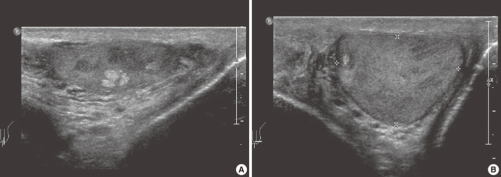
Fig. 1 Ultrasonography of the left testis. (A) A 0.4 × 0.5 × 0.4 cm sized poorly defined, heterogeneous mass with increased echogenicity. (B) Hypoechoic ring identified around the mass forming a "tubule-in-a-tubule" appearance.
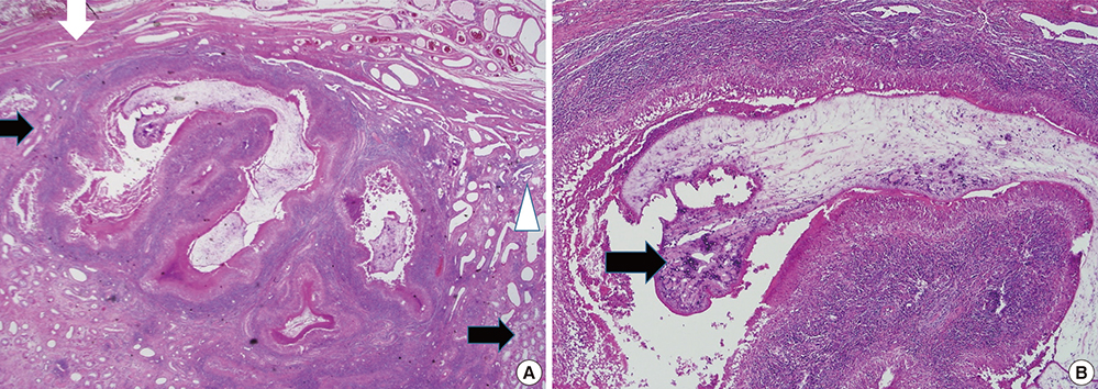
Fig. 2 Histopathological findings of the left testis. (A) A sparganum surrounded by abscess and necrotic materials is present within the testicular parenchyma. White arrows indicate seminiferous tubules, a black arrow indicates tunica albuginea, and a white triangle rete testis (×12.5). (B) The sparganum shows tortous recess of the bothrium, tegument, longitudinal layer of the smooth muscle, and loose internal stroma and multiple calcarous bodies are present in the loose internal stroma (×40).
Cited by 1 articles
-
Human Sparganosis in Korea
Jeong-Geun Kim, Chun-Seob Ahn, Woon-Mok Sohn, Yukifumi Nawa, Yoon Kong
J Korean Med Sci. 2018;33(44):. doi: 10.3346/jkms.2018.33.e273.
Reference
-
1. Nithiuthai S, Anantaphruti MT, Waikagul J, Gajadhar A. Waterborne zoonotic helminthiases. Vet Parasitol. 2004; 126:167–193.2. Kim DS, Park HJ, Kim SY, Kim SI, Lee TY, Park MH. Sparganosis in the testis. Korean J Urol. 2001; 42:883–885.3. Lee SY, Bae IH, Han GS, Cha SH, Kim SJ, Park KS, Song HK. A case of testis sparganosis. J Korean Soc Med Ultrasound. 2004; 23:147–150.4. Sakamoto T, Gutierrez C, Rodriguez A, Sauto S. Testicular sparganosis in a child from Uruguay. Acta Trop. 2003; 88:83–86.5. Lho YS, Kim HS, Yang SK. A case of sparganosis of the scrotum. Korean J Urol. 2001; 42:263–264.6. Sohn WM, Hong ST, Chai JY, Lee SH. Infectivity of the sparganum treated by praziquantel, gamma-irradiation and mechanical cutting. Korean J Parasitol. 1993; 31:135–139.7. Lee MK, Hong SJ, Kim HR. Seroprevalence of tissue invading parasitic infections diagnosed by ELISA in Korea. J Korean Med Sci. 2010; 25:1272–1276.
- Full Text Links
-
- Actions
-
Cited
- CITED
-
- Close
- Share
-
- Similar articles
-
- An Unusual Case of Testicular Seminoma Mimicking Segmental Testicular Infarction
- Testicular Tumor in Childhood: Report of 5 Cases
- A Case of Fibrous Pseudotumor of Testicular Tunic
- Diagnostic Value of Color Doppler Ultrasonography and Testicular Scintigraphy in Acute Scrotum
- A Case of Antenatal Testicular Torsion Mimicking Inflammation and Tumor in the Neonate
