A reliable method for evaluating upper molar distalization: Superimposition of three-dimensional digital models
- Affiliations
-
- 1Department of Orthodontics, Faculty of Dentistry, Karadeniz Technical University, Trabzon, Turkey.
- 2Department of Orthodontics, Faculty of Dentistry, Kocaeli University, Kocaeli, Turkey. burcuk12@yahoo.com
- 3Department of Orthodontics, Faculty of Dentistry, Gaziosmanpasa University, Tokat, Turkey.
- 4Department of Orthodontics, Faculty of Dentistry, Pamukkale University, Denizli, Turkey.
- KMID: 1787609
- DOI: http://doi.org/10.4041/kjod.2015.45.2.82
Abstract
OBJECTIVE
The aim of this study was to evaluate the reliability of measurements obtained after the superimposition of three-dimensional (3D) digital models by comparing them with those obtained from lateral cephalometric radiographs and photocopies of plaster models for the evaluation of upper molar distalization.
METHODS
Data were collected from plaster models and lateral cephalometric radiographs of 20 Class II patients whose maxillary first molars were distalized with an intraoral distalizer. The posterior movements of the maxillary first molars were evaluated using lateral cephalometric radiographs (group CP), photocopies of plaster models (group PH), and digitized 3D models (group TD). Additionally, distalization and expansion of the other teeth and the degrees of molar rotation were measured in group PH and group TD and compared between the two groups.
RESULTS
No significant difference was observed regarding the amount of molar distalization among the three groups. A comparison of the aforementioned parameters between group PH and group TD did not reveal any significant difference.
CONCLUSIONS
3D digital models are reliable to assess the results of upper molar distalization and can be considered a valid alternative to conventional measurement methods.
Keyword
MeSH Terms
Figure
-
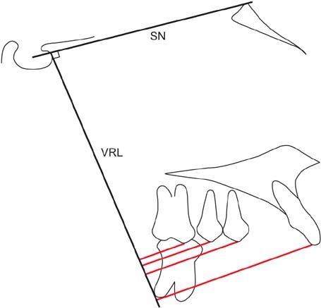
Figure 1 Cephalometric tracing and measurements. SN: Sella-nasion line, VRL: vertical reference line. Red lines: distalization amounts of the central incisor, premolar, and molar teeth.

Figure 2 Photocopy of a study model and the measured parameters (a, molar distalization; b, molar rotation; c, amount of expansion). Of the registration points, 1 is the most anterior point and 2 is the most posterior point of the incisive papilla.

Figure 3 Using three points as references, pre- (purple) and post-treatment (yellow) three-dimensional digital models were superimposed. Note that these points are not in the same plane, which provides a reliable and reproducible superimposition.

Figure 4 Measurements of molar distalization (a), molar rotation (b), and the amount of expansion (c) of the canines, premolars, and molars on a three-dimensional digital model.
Cited by 2 articles
-
Comparison of transverse dental changes induced by the palatally applied Frog appliance and buccally applied Karad's integrated distalizing system
Fatma Deniz Uzuner, Emine Kaygisiz, Fatih Unver, Tuba Tortop
Korean J Orthod. 2016;46(2):96-103. doi: 10.4041/kjod.2016.46.2.96.Cone-beam computed tomography-guided three-dimensional evaluation of treatment effectiveness of the Frog appliance
Mujia Li, Xiaoxia Su, Yang Li, Xianglin Li, Xinqin Si
Korean J Orthod. 2019;49(3):161-169. doi: 10.4041/kjod.2019.49.3.161.
Reference
-
1. Leifert MF, Leifert MM, Efstratiadis SS, Cangialosi TJ. Comparison of space analysis evaluations with digital models and plaster dental casts. Am J Orthod Dentofacial Orthop. 2009; 136:16.e1–16.e4.
Article2. Quimby ML, Vig KW, Rashid RG, Firestone AR. The accuracy and reliability of measurements made on computer-based digital models. Angle Orthod. 2004; 74:298–303.3. Stevens DR, Flores-Mir C, Nebbe B, Raboud DW, Heo G, Major PW. Validity, reliability, and reproducibility of plaster vs digital study models: comparison of peer assessment rating and Bolton analysis and their constituent measurements. Am J Orthod Dentofacial Orthop. 2006; 129:794–803.
Article4. Horton HM, Miller JR, Gaillard PR, Larson BE. Technique comparison for efficient orthodontic tooth measurements using digital models. Angle Orthod. 2010; 80:254–261.
Article5. Fleming PS, Marinho V, Johal A. Orthodontic measurements on digital study models compared with plaster models: a systematic review. Orthod Craniofac Res. 2011; 14:1–16.
Article6. Sjögren AP, Lindgren JE, Huggare JA. Orthodontic study cast analysis--reproducibility of recordings and agreement between conventional and 3D virtual measurements. J Digit Imaging. 2010; 23:482–492.
Article7. Zilberman O, Huggare JA, Parikakis KA. Evaluation of the validity of tooth size and arch width measurements using conventional and three-dimensional virtual orthodontic models. Angle Orthod. 2003; 73:301–306.8. Santoro M, Galkin S, Teredesai M, Nicolay OF, Cangialosi TJ. Comparison of measurements made on digital and plaster models. Am J Orthod Dentofacial Orthop. 2003; 124:101–105.
Article9. Gracco A, Buranello M, Cozzani M, Siciliani G. Digital and plaster models: a comparison of measurements and times. Prog Orthod. 2007; 8:252–259.10. Nalcaci R, Bicakci AA, Ozan F. Noncompliance screw supported maxillary molar distalization in a parallel manner. Korean J Orthod. 2010; 40:250–259.
Article11. Kim SJ, Chun YS, Jung SH, Park SH. Three dimensional analysis of tooth movement using different types of maxillary molar distalization appliances. Korean J Orthod. 2008; 38:376–387.
Article12. Ashmore JL, Kurland BF, King GJ, Wheeler TT, Ghafari J, Ramsay DS. A 3-dimensional analysis of molar movement during headgear treatment. Am J Orthod Dentofacial Orthop. 2002; 121:18–29.
Article13. Björk A, Skieller V. Normal and abnormal growth of the mandible. A synthesis of longitudinal cephalometric implant studies over a period of 25 years. Eur J Orthod. 1983; 5:1–46.
Article14. Champagne M. Reliability of measurements from photocopies of study models. J Clin Orthod. 1992; 26:648–650.15. Jang I, Tanaka M, Koga Y, Iijima S, Yozgatian JH, Cha BK, et al. A novel method for the assessment of three-dimensional tooth movement during orthodontic treatment. Angle Orthod. 2009; 79:447–453.
Article16. Lai EH, Yao CC, Chang JZ, Chen I, Chen YJ. Three-dimensional dental model analysis of treatment outcomes for protrusive maxillary dentition: comparison of headgear, miniscrew, and miniplate skeletal anchorage. Am J Orthod Dentofacial Orthop. 2008; 134:636–645.
Article17. Cha BK, Lee JY, Jost-Brinkmann PG, Yoshida N. Analysis of tooth movement in extraction cases using three-dimensional reverse engineering technology. Eur J Orthod. 2007; 29:325–331.
Article18. McGuinness NJ, Stephens CD. Storage of orthodontic study models in hospital units in the U.K. Br J Orthod. 1992; 19:227–232.
Article19. Thiruvenkatachari B, Al-Abdallah M, Akram NC, Sandler J, O'Brien K. Measuring 3-dimensional tooth movement with a 3-dimensional surface laser scanner. Am J Orthod Dentofacial Orthop. 2009; 135:480–485.
Article20. Kuroda T, Motohashi N, Tominaga R, Iwata K. Three-dimensional dental cast analyzing system using laser scanning. Am J Orthod Dentofacial Orthop. 1996; 110:365–369.
Article21. Hajeer MY, Millett DT, Ayoub AF, Siebert JP. Applications of 3D imaging in orthodontics: part I. J Orthod. 2004; 31:62–70.22. Mavropoulos A, Karamouzos A, Kiliaridis S, Papadopoulos MA. Efficiency of noncompliance simultaneous first and second upper molar distalization: a three-dimensional tooth movement analysis. Angle Orthod. 2005; 75:532–539.23. Bell A, Ayoub AF, Siebert P. Assessment of the accuracy of a three-dimensional imaging system for archiving dental study models. J Orthod. 2003; 30:219–223.
Article24. Keating AP, Knox J, Bibb R, Zhurov AI. A comparison of plaster, digital and reconstructed study model accuracy. J Orthod. 2008; 35:191–201.
Article25. Proffit WR, Fields HW, Sarver DM. Orthodontic diagnosis: the development of a problem list. In : Proffit WR, Fields HW, Sarver DM, editors. Contemporary orthodontics. 4th ed. St. Louis, Mo: Mosby Elsevier;2007. p. 167–233.26. Choi JI, Cha BK, Jost-Brinkmann PG, Choi DS, Jang IS. Validity of palatal superimposition of 3-dimensional digital models in cases treated with rapid maxillary expansion and maxillary protraction headgear. Korean J Orthod. 2012; 42:235–241.
Article27. Bailey LT, Esmailnejad A, Almeida MA. Stability of the palatal rugae as landmarks for analysis of dental casts in extraction and nonextraction cases. Angle Orthod. 1996; 66:73–78.28. Hoggan BR, Sadowsky C. The use of palatal rugae for the assessment of anteroposterior tooth movements. Am J Orthod Dentofacial Orthop. 2001; 119:482–488.
Article29. Simmons JD, Moore RN, Erickson LC. A longitudinal study of anteroposterior growth changes in the palatine rugae. J Dent Res. 1987; 66:1512–1515.
Article30. Choi DS, Jeong YM, Jang I, Jost-Brinkmann PG, Cha BK. Accuracy and reliability of palatal superimposition of three-dimensional digital models. Angle Orthod. 2010; 80:497–503.
Article
- Full Text Links
-
- Actions
-
Cited
- CITED
-
- Close
- Share
-
- Similar articles
-
- Validity of palatal superimposition of 3-dimensional digital models in cases treated with rapid maxillary expansion and maxillary protraction headgear
- Noncompliance screw supported maxillary molar distalization in a parallel manner
- The frog appliance for upper molar distalization: a case report
- Zygoma-gear appliance for intraoral upper molar distalization
- A method for mandibular dental arch superimposition using 3D cone beam CT and orthodontic 3D digital model
