J Korean Med Sci.
2013 Mar;28(3):383-387. 10.3346/jkms.2013.28.3.383.
Bronchial Anthracofibrosis and Macroscopic Tissue Pigmentation on EBUS-TBNA Predict a Low Probability of Metastatic Lymphadenopathy in Korean Lung Cancer Patients
- Affiliations
-
- 1Department of Pulmonary and Critical Care Medicine, Asan Medical Center, University of Ulsan College of Medicine, Seoul, Korea. ccm@amc.seoul.kr
- 2Department of Oncology, Asan Medical Center, University of Ulsan College of Medicine, Seoul, Korea.
- KMID: 1786935
- DOI: http://doi.org/10.3346/jkms.2013.28.3.383
Abstract
- The identification of mediastinal lymph nodes (LNs) in lung cancer is an important step of treatment decision and prognosis prediction. The endobronchial ultrasound-guided transbronchial needle aspiration (EBUS-TBNA) is widely used to assess the mediastinal LNs and tissue confirmation in lung cancer. As use of bronchoscopy or EBUS-TBNA has been increased, bronchial anthracofibrosis (BAF) has been detected frequently. Moreover, BAF is often accompanied by mediastinal lymphadenopathy and showed false-positive positron emission tomography uptake, which mimics metastatic lymphadenopathy in lung cancer patients. However, clinical implication of BAF during bronchoscopy is not well understood in lung cancer. We retrospectively reviewed 536 lung cancer patients who performed EBUS-TBNA and observed BAF in 55 patients. A total of 790 LNs were analyzed and macroscopic tissue pigmentation was observed in 228 patients. The adjusted odds ratio for predicting malignant LN was 0.46 for BAF, and 0.22 for macroscopic tissue pigmentation. The specificity of BAF and macroscopic tissue pigmentation for predicting a malignant LN was 75.7% and 42.2%, respectively, which was higher than the specificity of using LN size or standard uptake value on PET. In conclusion, BAF and macroscopic tissue pigmentation during EBUS-TBNA are less commonly found in malignant LNs than reactive LNs in Korean lung cancer patients.
MeSH Terms
-
Adult
Aged
Aged, 80 and over
Asian Continental Ancestry Group
Biopsy, Fine-Needle
Bronchi/*pathology
Bronchoscopy
Carcinoma, Non-Small-Cell Lung/*pathology/radiography
Constriction, Pathologic
Female
Humans
Logistic Models
Lung Neoplasms/*pathology/radiography
Lymph Nodes/pathology
Lymphatic Diseases/*pathology
Lymphatic Metastasis
Male
Middle Aged
Odds Ratio
Pigmentation
Positron-Emission Tomography
Predictive Value of Tests
Republic of Korea
Retrospective Studies
Small Cell Lung Carcinoma/*pathology/radiography
Ultrasonography, Interventional
Figure
-
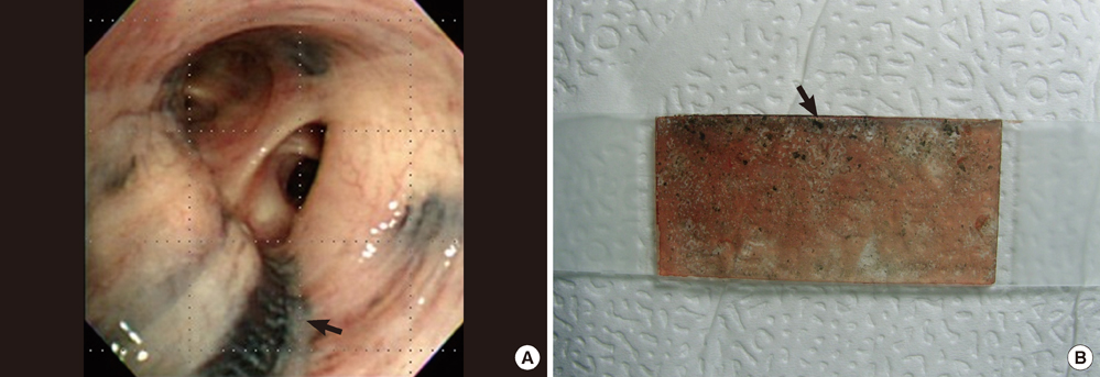
Fig. 1 Endobronchial findings. (A) Bronchial anthracofibrosis. (B) Macroscopic tissue pigmentation observed during EBUS-TBNA.
Reference
-
1. Ettinger DS, Akerley W, Bepler G, Blum MG, Chang A, Cheney RT, Chirieac LR, D'Amico TA, Demmy TL, Ganti AK, et al. Non-small cell lung cancer. J Natl Compr Canc Netw. 2010. 8:740–801.2. Lee BE, Kletsman E, Rutledge JR, Korst RJ. Utility of endobronchial ultrasound-guided mediastinal lymph node biopsy in patients with non-small cell lung cancer. J Thorac Cardiovasc Surg. 2012. 143:585–590.3. Gupta A, Shah A. Bronchial anthracofibrosis: an emerging pulmonary disease due to biomass fuel exposure. Int J Tuberc Lung Dis. 2011. 15:602–612.4. Kim MA, Lee JC, Choi C. False-positive FDG-PET and bronchial anthracofibrosis. J Thorac Oncol. 2012. 7:1474.5. Jang SJ, Lee SY, Kim SC, Lee SY, Cho HS, Park KH, Moon HS, Song JS, Park SH, Kim YK, et al. Clinical and radiological characteristics of non-tuberculous bronchial anthracofibrosis. Tuberc Respir Dis. 2007. 63:139–144.6. Kim HY, Im JG, Goo JM, Kim JY, Han SK, Lee JK, Song JW. Bronchial anthracofibrosis (inflammatory bronchial stenosis with anthracotic pigmentation): CT findings. AJR Am J Roentgenol. 2000. 174:523–527.7. Bilici A, Erdem T, Boysan SN, Acbay O, Oz B, Besirli K, Gundogdu S. A case of anthracosis presenting with mediastinal lymph nodes mimicking tuberculous lymphadenitis or malignancy. Eur J Intern Med. 2003. 14:444–446.8. Kim YK, Lee KS, Kim BT, Choi JY, Kim H, Kwon OJ, Shim YM, Yi CA, Kim HY, Chung MJ. Mediastinal nodal staging of nonsmall cell lung cancer using integrated 18F-FDG PET/CT in a tuberculosis-endemic country: diagnostic efficacy in 674 patients. Cancer. 2007. 109:1068–1077.9. Konishi J, Yamazaki K, Tsukamoto E, Tamaki N, Onodera Y, Otake T, Morikawa T, Kinoshita I, Dosaka-Akita H, Nishimura M. Mediastinal lymph node staging by FDG-PET in patients with non-small cell lung cancer: analysis of false-positive FDG-PET findings. Respiration. 2003. 70:500–506.10. Lee SH, Min JW, Lee CH, Park CM, Goo JM, Chung DH, Kang CH, Kim YT, Kim YW, Han SK, et al. Impact of parenchymal tuberculosis sequelae on mediastinal lymph node staging in patients with lung cancer. J Korean Med Sci. 2011. 26:67–70.11. Choi HY, Kim YK, Lee JJ, Kim SE. Bronchial anthracofibrosis: a potential false-positive finding on F-18 FDG PET. Ann Nucl Med. 2012. 26:681–683.12. Fujiwara T, Yasufuku K, Nakajima T, Chiyo M, Yoshida S, Suzuki M, Shibuya K, Hiroshima K, Nakatani Y, Yoshino I. The utility of sonographic features during endobronchial ultrasound-guided transbronchial needle aspiration for lymph node staging in patients with lung cancer: a standard endobronchial ultrasound image classification system. Chest. 2010. 138:641–647.13. Nguyen P, Bashirzadeh F, Hundloe J, Salvado O, Dowson N, Ware R, Masters IB, Bhatt M, Kumar AR, Fielding D. Optical differentiation between malignant and benign lymphadenopathy by grey scale texture analysis of endobronchial ultrasound convex probe images. Chest. 2012. 141:709–715.14. Silvestri GA, Tanoue LT, Margolis ML, Barker J, Detterbeck F. American College of Chest Physicians. The noninvasive staging of non-small cell lung cancer: the guidelines. Chest. 2003. 123:147S–156S.
- Full Text Links
-
- Actions
-
Cited
- CITED
-
- Close
- Share
-
- Similar articles
-
- Paraesophageal Anthracofibrosis Mimicking Metastatic Lymphadenopathy in Papillary Thyroid Cancer: a Case Report
- The First Pediatric Case of Intrathoracic Tuberculosis Lymphadenitis Diagnosed by Endobronchial Ultrasound Guided Transbronchial Needle Aspiration
- Bronchial Bleeding Induced by Suction in a Patient with Bronchial Anthracofibrosis
- A case of renal cell carcinoma with mediastinal lymph node metastasis diagnosed by EBUS-TBNA
- Usefulness of Endobronchial Ultrasound-Guided Transbronchial Needle Aspiration for Diagnosis of Sarcoidosis
