Surgical Treatment of Congenital Hallux Varus
- Affiliations
-
- 1Department of Orthopaedic Surgery, Samsung Medical Center, Sungkyunkwan University School of Medicine, Seoul, Korea.
- 2Department of Orthopaedic Surgery, Eulji Hospital, Eulji University School of Medicine, Seoul, Korea. fromspace@hanmail.net
- 3Department of Orthopaedic Surgery, Ilsan Paik Hospital, Inje University School of Medicine, Goyang, Korea.
- KMID: 1784670
- DOI: http://doi.org/10.4055/cios.2014.6.2.216
Abstract
- BACKGROUND
The purpose of this study was to report outcomes of congenital hallux varus deformity after surgical treatment.
METHODS
We evaluated ten feet of eight patients with a congenital hallux varus deformity, including four feet combined with a longitudinal epiphyseal bracket (LEB). There were seven male patients and one female patient with a mean age of 33 months (range, 7 to 103 months) at the time of surgery. Two patients were bilaterally involved. The mean duration of follow-up was 5.9 years (range, 2.3 to 13.8 years). Clinical outcomes were assessed according to the criteria of Phelps and Grogan. Surgical procedures included the Farmer procedure, the McElvenny procedure or an osteotomy at the first metatarsal or proximal phalanx.
RESULTS
The clinical results were excellent in two feet, good in six and poor in two feet. The LEB was associated with hallux varus in four feet and were treated by osteotomy alone or in conjunction with soft tissue procedure.
CONCLUSIONS
Congenital hallux varus was successfully corrected by surgery with overall favorable outcome. Preoperatively, a LEB should be considered as a possible cause of the deformity in order to prevent recurrent or residual varus after surgery.
MeSH Terms
Figure
-
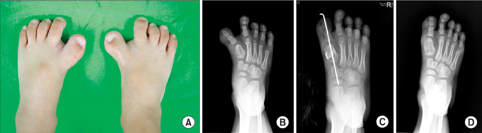
Fig. 1 Patient 7. (A) Preoperative photograph showing bilateral hallux varus with widening of the first web space. Preoperative scars due to removal of accessory toes are also noted. (B) Preoperative radiograph of the right foot at 58 months of age showing a short thickened first metatarsal, which might result from closure of the physis between the bracket and diaphysis. Radiographs at the immediate postoperative follow-up (C) and at the final follow-up (D).
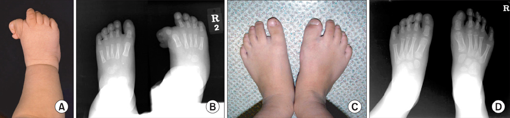
Fig. 2 Patient 2. (A) Preoperative photograph showing marked medial deviation of the broad great toe and widening of the first web space. (B) Preoperative radiograph showing varus angulation of the first metatarsophalangeal joint and accessory bone of the great toe. (C) Follow-up photograph. (D) Sufficient correction with cosmetically satisfactory appearance of the foot was observed at four years after the Farmer technique and medial open wedge osteotomy at the proximal phalanx. The final result was graded as excellent at 10 years after surgery.
Reference
-
1. Farmer AW. Congenital hallux varus. Am J Surg. 1958; 95(2):274–278.2. Herring JA. Tachdjian's pediatric orthopaedics. 4th ed. Philadelphia, PA: Saunders;2008. p. 1172–1173.3. Light TR, Ogden JA. The longitudinal epiphyseal bracket: implications for surgical correction. J Pediatr Orthop. 1981; 1(3):299–305.4. Masada K, Tsuyuguchi Y, Kawabata H, Ono K. Treatment of preaxial polydactyly of the foot. Plast Reconstr Surg. 1987; 79(2):251–258.5. Mills JA, Menelaus MB. Hallux varus. J Bone Joint Surg Br. 1989; 71(3):437–440.6. Mubarak SJ, O'Brien TJ, Davids JR. Metatarsal epiphyseal bracket: treatment by central physiolysis. J Pediatr Orthop. 1993; 13(1):5–8.7. Phelps DA, Grogan DP. Polydactyly of the foot. J Pediatr Orthop. 1985; 5(4):446–451.8. Glickman SH, Cornfield RH. Surgical reconstruction of a congenital foot deformity: hallux varus with brachymetatarsia of the first metatarsal. J Foot Surg. 1990; 29(5):499–503.9. Joseph B, Jacob T, Chacko V. Hallux varus: a study of thirty cases. J Foot Surg. 1984; 23(5):392–397.10. Stanifer E, Hodor D, Wertheimer S. Congenital hallux varus: case presentation and review of the literature. J Foot Surg. 1991; 30(5):509–512.11. McElvenny RT. Hallux varus. Q Bull Northwest Univ Med Sch. 1941; 15:277–280.12. Thomson SA. Hallux varus and metatarsus varus: a five-year study (1954-1958). Clin Orthop. 1960; 16:109–118.13. Sobel E, Levitz S, Cohen R, Giorgini R, Jules KT. Longitudinal epiphyseal bracket: associated foot deformities with implications for treatment. J Am Podiatr Med Assoc. 1996; 86(4):147–155.14. Mahboubi S, Davidson R. MR imaging in longitudinal epiphyseal bracket in children. Pediatr Radiol. 1999; 29(4):259–261.15. Shea KG, Mubarak SJ, Alamin T. Preossified longitudinal epiphyseal bracket of the foot: treatment by partial bracket excision before ossification. J Pediatr Orthop. 2001; 21(3):360–365.16. Kucukkaya M, Kabukcuoglu Y, Tezer M, Kuzgun U. Correcting and lengthening of metatarsal deformity with circular fixator by distraction osteotomy: a case of longitudinal epiphyseal bracket. Foot Ankle Int. 2002; 23(5):427–432.17. Marcdargent Fassier A, Gueffier X, Fraisse T, Janelle C, Fassier F. Longitudinal epiphyseal bracket of the first metatarsus (delta bone). Rev Chir Orthop Reparatrice Appar Mot. 2007; 93(5):486–493.18. Morrissy RT, Weinstein SL. Lovell and Winter's pediatric orthopaedics. 6th ed. Philadelphia, PA: Lippincott Williams & Wilkins;2005. p. 1306.19. Schreck MA. Pediatric longitudinal epiphyseal bracket: review and case presentation. J Foot Ankle Surg. 2006; 45(5):342–345.20. Lampropulos M, Puigdevall M, Zapozko D, Malvarez H. Treatment of first metatarsal longitudinal epiphyseal bracket by excision before closure. J Foot Ankle Surg. 2007; 46(4):297–301.21. Mubarak SJ, O'Brien TJ, Davids JR. Metatarsal epiphyseal bracket: treatment by central physiolysis. J Pediatr Orthop. 1993; 13(1):5–8.
- Full Text Links
-
- Actions
-
Cited
- CITED
-
- Close
- Share
-
- Similar articles
-
- Operative Treatment of the Bilateral 1,4th Brachymetatarsia with Painful Callosity and Hallux Varus using Massive Metatarsal Axial Shortening: A Case Report
- Minimally Invasive Surgery with Tenorrhaphy for Postoperative Hallux Varus Deformity Combined with Flexor Hallucis Longus Rupture after Hallux Valgus Correction: A Case Report
- Complications after Surgical Correction of Hallux Valgus
- Tibial Torsion and Knee Varus Angle: Are These Aetiologies of Hallux Valgus?
- Patient Characteristics and Treatment of Hallux Polydactyly Associated with Varus Deformity
